. 2021 Mar 17:12:655239.
doi: 10.3389/fmicb.2021.655239. eCollection 2021.

Interdependent Polar Localization of FlhF and FlhG and Their Importance for Flagellum Formation of Vibrio parahaemolyticus

Affiliations

Interdependent Polar Localization of FlhF and FlhG and Their Importance for Flagellum Formation of Vibrio parahaemolyticus

Erick Eligio Arroyo-Pérez et al. Front Microbiol. .

Abstract

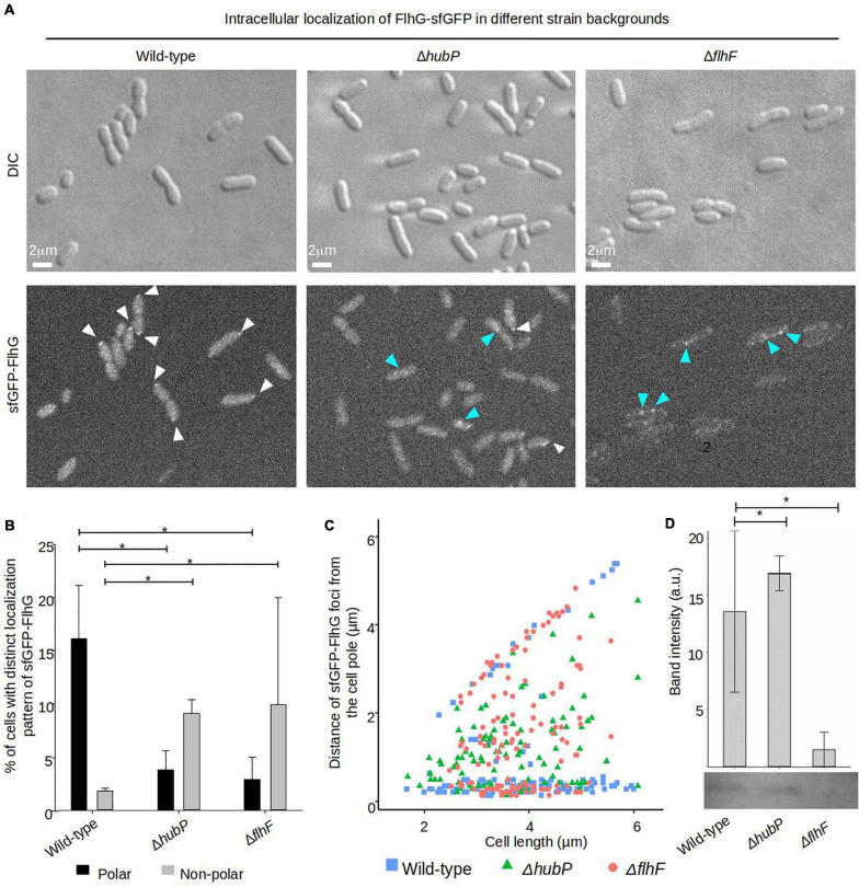
Failure of the cell to properly regulate the number and intracellular positioning of their flagella, has detrimental effects on the cells' swimming ability. The flagellation pattern of numerous bacteria is regulated by the NTPases FlhF and FlhG. In general, FlhG controls the number of flagella produced, whereas FlhF coordinates the position of the flagella. In the human pathogen Vibrio parahaemolyticus, its single flagellum is positioned and formed at the old cell pole. Here, we describe the spatiotemporal localization of FlhF and FlhG in V. parahaemolyticus and their effect on swimming motility. Absence of either FlhF or FlhG caused a significant defect in swimming ability, resulting in absence of flagella in a ΔflhF mutant and an aberrant flagellated phenotype in ΔflhG. Both proteins localized to the cell pole in a cell cycle-dependent manner, but displayed different patterns of localization throughout the cell cycle. FlhF transitioned from a uni- to bi-polar localization, as observed in other polarly flagellated bacteria. Localization of FlhG was strictly dependent on the cell pole-determinant HubP, while polar localization of FlhF was HubP independent. Furthermore, localization of FlhF and FlhG was interdependent and required for each other's proper intracellular localization and recruitment to the cell pole. In the absence of HubP or FlhF, FlhG forms non-polar foci in the cytoplasm of the cell, suggesting the possibility of a secondary localization site within the cell besides its recruitment to the cell poles.

Keywords: FlhF; FlhG; HubP; Vibrio parahaemolyticus; flagellum; intracellular organization.


Conflict of interest statement

The authors declare that the research was conducted in the absence of any commercial or financial relationships that could be construed as a potential conflict of interest.

Figures

FIGURE 1
FIGURE 1
FlhF and FlhG regulate swimming and flagellum production in V. parahaemolyticus. (A) Representative image of a swimming assay in soft agar of indicated V. parahaemolyticus strains. (B) Bar graph showing the average diameter of swimming colonies of the indicated V. parahaemolyticus strains relative to wild-type cells. (C) Representative transmission electron micrographs of the indicated V. parahaemolyticus strains stained with uranyl acetate. (D) Bar graph depicting the average percentage of cells with distinct flagellation patterns, n = 200 cells. (B,D) Asterisk, *, indicates p < 0.05, Tested with ANOVA in blocks + Tukey HSD. Error bars indicate standard deviation.
FIGURE 2
FIGURE 2
The intracellular localization of FlhF and FlhG in V. parahaemolyticus. (A) DIC and fluorescence microscopy of V. parahaemolyticus strains expressing FlhF-sfGFP or sfGFP-FlhG fusion proteins. White arrows indicate polar foci, orange arrows = unipolar foci, green arrows = bipolar foci, red arrow = diffuse. (B) Bar graph showing the percentages of cells with fluorescent foci at one, two, or no poles. Asterisk, *, indicates p < 0.05, tested with ANOVA + Tukey HSD. (C) Graph depicting the distance of FlhF-sfGFP clusters from the cell poles as a function of cell 2length.
FIGURE 3
FIGURE 3
A dynamic spatiotemporal intracellular localization of FlhF and FlhG during the V. parahaemolyticus cell cycle. (A,B) Time-lapse DIC and fluorescence microscopy of V. parahaemolyticus strains expressing (A) FlhF-sfGFP or (B) sfGFP-FlhG fusion proteins. White numbers indicate minutes elapsed.
FIGURE 4
FIGURE 4
FlhG is required for proper intracellular localization of FlhF. (A) DIC and fluorescence microscopy of indicated V. parahaemolyticus strains expressing FlhF-sfGFP. White arrows indicate polar FlhF-sfGFP foci. (B) Demographs showing the fluorescence intensity of sfGFP along the cell length in a population of V. parahaemolyticus cells relative to cell length. Demographs include date from >600 cells pooled from three distinct experiments. (C) Bar graph showing the percentage of cells with distinct FlhF-sfGFP localization patterns in the indicated V. parahaemolyticus strain backgrounds. Asterisk, *, indicates p < 0.05, tested with ANOVA in blocks + Tukey HSD. Error bars indicate standard deviation. (D) Box plot showing the fluorescence intensity of polar FlhF-sfGFP foci of the indicated V. parahaemolyticus strains. Asterisk, *, indicates p < 0.05, tested with ANOVA + Tukey HSD. (E) Western blot with anti-GFP monoclonal antibody against whole cell extract of strains expressing FlhF-sfGFP. The bar-graph depicts the quantification of the signal detected from three biological replicates. Error bars indicate standard deviation and asterisk, *, indicates p < 0.05, tested with student’s t-test.
FIGURE 5
FIGURE 5
Proper intracellular localization of FlhG is regulated by HubP and FlhF. (A) DIC and fluorescence microscopy of sfGFP-FlhG in the indicated V. parahaemolyticus strains. White arrows indicate polar foci of sfGFP-FlhG and blue arrows indicate cytoplasmic clusters. (B) Bar graphs showing the percentage of cells with distinct localization pattern of sfGFP-FlhG in the indicated strains of V. parahaemolyticus. Error bars indicate standard deviation and asterisk, *, indicates p < 0.05, tested with ANOVA + Tukey HSD. (C) Graph depicting the distance of sfGFP-FlhG clusters from the cell poles as a function of cell length in the indicated V. parahaemolyticus strain backgrounds. (D) Western blot with anti-GFP monoclonal antibody against whole cell extract of strains expressing sfGFP-FlhG. The bar-graph depicts the quantification of the signal detected from three biological replicates. Error bars indicate standard deviation and asterisk, *, indicates p < 0.05, tested with student’s t-test.

References

    1. Alvarado A., Kjær A., Yang W., Mann P., Briegel A., Waldor M. K., et al. (2017). Coupling chemosensory array formation and localization. Elife 6:e31058. 10.7554/eLife.31058 - DOI - PMC - PubMed
    1. Balaban M., Joslin S. N., Hendrixson D. R. (2009). FlhF and its GTPase activity are required for distinct processes in flagellar gene regulation and biosynthesis in Campylobacter jejuni. J. Bacteriol. 191 6602–6611. 10.1128/JB.00884-09 - DOI - PMC - PubMed
    1. Bange G., Kümmerer N., Grudnik P., Lindner R., Petzold G., Kressler D., et al. (2011). Structural basis for the molecular evolution of SRP-GTpase activation by protein. Nat. Struct. Mol. Biol. 18 1376–1380. 10.1038/nsmb.2141 - DOI - PubMed
    1. Baumann P., Baumann L. (1977). Biology of the marine enterobacteria: genera Beneckea and Photobacterium. Annu. Rev. Microbiol. 31 39–61. 10.1146/annurev.mi.31.100177.000351 - DOI - PubMed
    1. Blagotinsek V., Schwan M., Steinchen W., Mrusek D., Hook J. C., Rossmann F., et al. (2020). An ATP-dependent partner switch links flagellar C-ring assembly with gene expression. Proc. Natl. Acad. Sci. U.S.A. 117 20826–20835. 10.1073/pnas.2006470117 - DOI - PMC - PubMed

LinkOut - more resources