. 2021 Mar 17:12:650621.
doi: 10.3389/fimmu.2021.650621. eCollection 2021.

Delayed Gut Colonization Shapes Future Allergic Responses in a Murine Model of Atopic Dermatitis

Affiliations

Delayed Gut Colonization Shapes Future Allergic Responses in a Murine Model of Atopic Dermatitis

Amalie W Arildsen et al. Front Immunol. .

Abstract

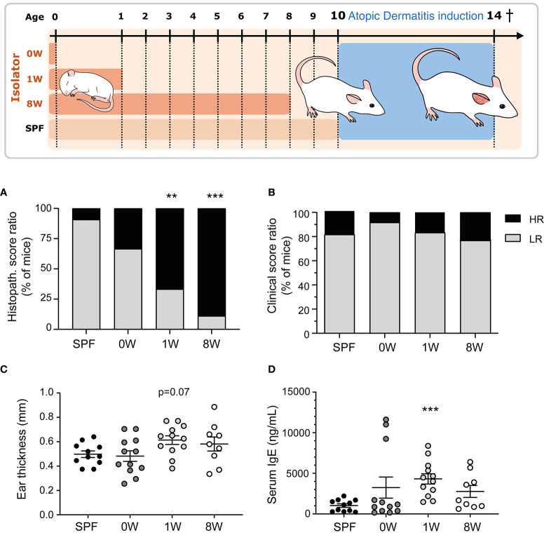
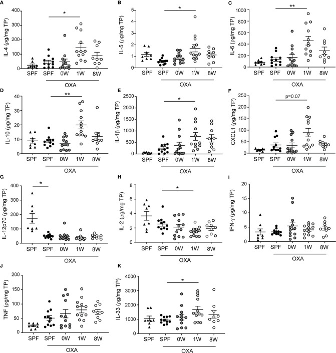
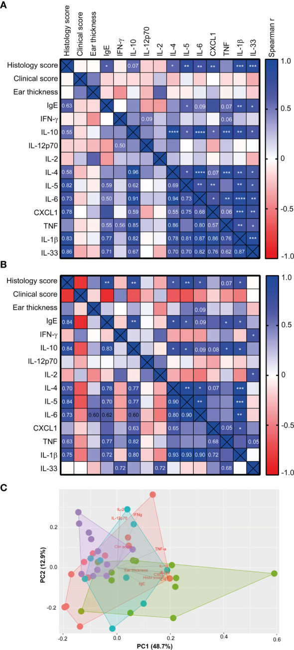
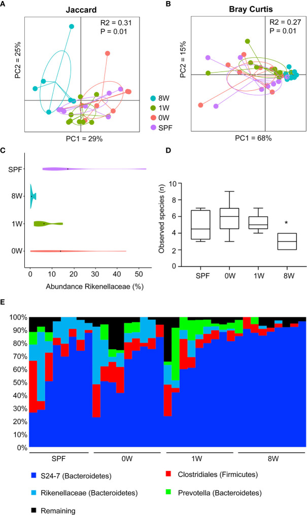
Epidemiological studies have long reported that perturbations of the childhood microbiome increase the risk of developing allergies, but a causal relationship with atopic dermatitis remains unclear. Here we colonized germ-free mice at birth or at one or eight week-of-age to investigate the role of prenatal and early postnatal microbial exposure on development of oxozolone-induced dermatitis later in life. We demonstrate that only one week delayed microbial colonization increased IgE levels and the total histological score of the inflamed ear compared to mice colonized throughout life. In parallel, several pro-inflammatory cytokines and chemokines were upregulated in the ear tissue demonstrating an enhanced immunological response following delayed postnatal colonization of the gut. In contrast, sensitivity to oxazolone-induced dermatitis was unaffected by the presence of a maternal microbiota during gestation. Mice colonized at eight week-of-age failed to colonize Rikenellaceae, a group of bacteria previously associated with a high-responding phenotype, and did not develop an immunological response to the same extent as the early colonized mice despite pronounced histopathological manifestations. The study provides proof-of-principle that the first intestinal colonizers of mice pups are crucial for the development of oxazolone-induced dermatitis later in life, and that the status of the maternal microbiota during pregnancy has no influence on the offspring's allergic immune response. This highlights an important window of opportunity following birth for microbiota-mediated interventions to prevent atopic responses later in life. How long such a window is open may vary between mice and humans considering species differences in the ontogeny of the immune system.

Keywords: allergy; atopic dermatitis; childhood ezcema; early priming; gut microbiota; immune maturation; rikenellaceae; window of opportunity.


Conflict of interest statement

The authors declare that the research was conducted in the absence of any commercial or financial relationships that could be construed as a potential conflict of interest.

Figures

Figure 1
Figure 1
Sensitivity to oxazolone-induced dermatitis in SPF and ex-germ-free mice. The experimental setup is outlined with three germ-free groups colonized at birth (0W, n=12), 1 week-of-age (1W, n=12), or 8 week-of-age (8W, n=9) and one microbiota-associated SPF bred control group (SPF, n=11). After 10 weeks-of-age, the mice were sensitized with 0.8% oxazolone before they were challenged ten times with 0.2% oxazolone solution on the right ear. The mice were euthanized at 14 weeks of age after the last challenge and sampled for further analyses. (A) HE stained cross sections of the inflamed ears were scored blindly in all mice. The percentages of mice expressing a high (total score >8.5) or low (total score ≤8.5) responding phenotype are shown. (B) The degree of oxazolone-induced dermatitis was clinically scored by two blinded persons independently in all mice when euthanized. The percentages of mice expressing a high (average total score >7.5) and low (average total score ≤7.5) phenotype are shown. (C) Ear thickness was measured twice on anaesthetized mice immediately before euthanization. Mean and SEM are shown. (D) Serum IgE was measured with ELISA in all mice. Mean and SEM are shown. ** indicate p < 0.01, *** indicate p < 0.001. All p -values below 0.1 are indicated.
Figure 2
Figure 2
Ear tissue cytokine levels were upregulated after a short-term delayed colonization. (A–K) Cytokines were measured with two mesoscale multiplex kit in punch biopsies sampled from the inflamed ear (SPF, n=11, 0W, n=12, 1W, n=12, 8W, n=9) and from untreated ears of SPF mice (n=6) for comparison. The levels of each cytokine and chemokine were normalized to the total protein concentration per sample. Mean and SEM are shown for each cytokine. * indicates p < 0.05, ** indicate p < 0.01. All p -values below 0.1 are indicated.
Figure 3
Figure 3
The histology score correlated with the immune response of high-responding mice Heatmaps illustrating significant correlations between phenotypic disease markers used to characterize disease development and immunological responses in the 1W group (n=12) (A), and in the 8W group (B). P-values are indicated in the upper right half of the heatmaps and the r2 values are given in the lower left halfs for correlations with p-values < 0.1. The color codes indicate the positive or negative r value (correlation). (C) Principal component analysis plot based on the phenotypic disease and immune markers listed in (A), showing clusters of SPF (n=11, purple), 0W (n=12, red), 1W (n=12, green) and 8W (n=9, blue) mice. * indicates p < 0.05, ** indicate p < 0.01, *** indicate p < 0.001, **** indicate p < 0.0001.
Figure 4
Figure 4
Late colonization alters the gut microbiota composition. Principal coordinates analysis plot of nanopore based sequencing of the near full 16S rRNA gene amplicon based on (A) Jaccard (unweighted) and (B) Bray-Curtis (weighted) distance matrices as indicated. The plots illustrate feces samples from SPF (n=8, purple), 0W (n=9, red), 1W (n=9, green) and 8W (n=9, blue) mice before sensitization with oxazolone at 10 week-of-age. (C) Violin plot illustrating the abundance of the only taxa significantly different between the groups, the family Rikenellaceae. (D) Alpha diversity plot showing the observed species index based on species level summarized ASV table. (E) Chart illustrating the distribution of the main taxa colonizing the gut. * indicates p < 0.05.

Similar articles

Cited by

References

    1. Al NZ, Eberl G. Imprinting of the immune system by the microbiota early in life. Mucosal Immunol (2020) 13(2):183–9. 10.1038/s41385-020-0257-y - DOI - PubMed
    1. Hansen CH, Nielsen DS, Kverka M, Zakostelska Z, Klimesova K, Hudcovic T, et al. . Patterns of early gut colonization shape future immune responses of the host. PLoS One (2012) 7(3):e34043. 10.1371/journal.pone.0034043 - DOI - PMC - PubMed
    1. Stokholm J, Blaser MJ, Thorsen J, Rasmussen MA, Waage J, Vinding RK, et al. . Maturation of the gut microbiome and risk of asthma in childhood. Nat Commun (2018) 9(1):141. 10.1038/s41467-017-02573-2 - DOI - PMC - PubMed
    1. Zimmermann P, Messina N, Mohn WW, Finlay BB, Curtis N. Association between the intestinal microbiota and allergic sensitization, eczema, and asthma: A systematic review. J Allergy Clin Immunol (2019) 143(2):467–85. 10.1016/j.jaci.2018.09.025 - DOI - PubMed
    1. Masoli M, Fabian D, Holt S, Beasley R. The global burden of asthma: executive summary of the GINA Dissemination Committee report. Allergy (2004) 59(5):469–78. 10.1111/j.1398-9995.2004.00526.x - DOI - PubMed

MeSH terms