Review
. 2021 Mar 18:12:655697.
doi: 10.3389/fimmu.2021.655697. eCollection 2021.

Hepatocellular Carcinoma Immune Landscape and the Potential of Immunotherapies

Affiliations
Review

Hepatocellular Carcinoma Immune Landscape and the Potential of Immunotherapies

Julie Giraud et al. Front Immunol. .

Abstract

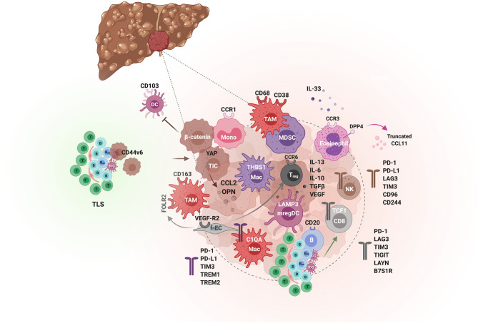
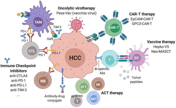
Hepatocellular carcinoma (HCC) is the most common liver tumor and among the deadliest cancers worldwide. Advanced HCC overall survival is meager and has not improved over the last decade despite approval of several tyrosine kinase inhibitors (TKi) for first and second-line treatments. The recent approval of immune checkpoint inhibitors (ICI) has revolutionized HCC palliative care. Unfortunately, the majority of HCC patients fail to respond to these therapies. Here, we elaborate on the immune landscapes of the normal and cirrhotic livers and of the unique HCC tumor microenvironment. We describe the molecular and immunological classifications of HCC, discuss the role of specific immune cell subsets in this cancer, with a focus on myeloid cells and pathways in anti-tumor immunity, tumor promotion and immune evasion. We also describe the challenges and opportunities of immunotherapies in HCC and discuss new avenues based on harnessing the anti-tumor activity of myeloid, NK and γδ T cells, vaccines, chimeric antigen receptors (CAR)-T or -NK cells, oncolytic viruses, and combination therapies.

Keywords: NASH; cirrhosis; immune checkpoint inhibitors; immunosuppression; immunotherapy; inflammation; tumor microenvironment; tumor-associated macrophages.


Conflict of interest statement

The authors declare that the research was conducted in the absence of any commercial or financial relationships that could be construed as a potential conflict of interest.

Figures

Figure 1
Figure 1
HCC etiologies, genetic predisposition and current standard of care for the advanced stage. (A) HCC etiologies include chronic infection with HBV or HCV, alcohol abuse, dietary toxins and/or the metabolic syndrome linked to obesity and type 2 diabetes. In rare cases, HCC stems from a monogenic disease e.g., hemochromatosis, caused by mutations in the homeostatic iron regulator gene HFE1; Wilson disease involving mutations in the ATPase copper transporting beta gene ATP7B; tyrosinemia, resulting from mutations in the gene encoding fumarylacetoacetate hydrolase FAH, α1-trypsin deficiency caused by mutations in serpin family A member 1 SERPINA1; or glycogen storage disease, in which the glucose-6-phosphatase gene is mutated. (B) The standard of care for treating patients with advanced HCC has been revised with the approval of immune checkpoint inhibitors. In first line, patients are administered TKi, mainly sorafenib or lenvatinib, or given the newly approved combination of bevacizumab (anti-VEGF) + atezolizumab (anti-PD-L1). In second line, patients refractory to TKi are treated with other TKIs, whereas anti-PD-1 ICI, nivolumab or pembrolizumab, have only been approved in the USA as an option for second line (despite the lack of superior efficacity in phase III trials compared to TKi).
Figure 2
Figure 2
Architecture of the human liver and its immune system. (A) Schematic illustration of the human liver anatomy namely its 8 segments, hepatic lobules, and triads of portal vein/hepatic artery/bile duct. (B) The liver zonation. Oxygen and metabolic gradients define three liver zones with specialized hepatocytes functions. (C) A zoom on hepatic cellular interactions across the sinusoids, the space of Disse and the hepatocyte plates. Liver sinusoidal endothelial cells (LSECs) line the liver sinusoid by forming a fenestrated monolayer. Their basal side interacts with hepatocytes and hepatic stellate cells (HSCs) in the space of Disse, whereas their luminal side interacts with liver-resident leukocytes, including Kupffer cells (KCs).
Figure 3
Figure 3
The landscapes of the normal, cirrhotic and HCC-bearing livers. All immune and non-immune cell types identified by high-resolution single cell analyses of the human healthy and cirrhotic livers and of HCC are illustrated along with their discriminatory markers. Arrows depict direction of change in cirrhosis or HCC vs. the normal liver, with green arrows indicating an expansion, red arrows a depletion and blue horizontal arrow no change in the examined cell subset. The cellular landscapes of the healthy and cirrhotic livers were from (26). The information on the HCC landscape was from (27), but with complementary information from the following studies: γδ T cells and M2 macrophages (28), cancer-associated fibroblasts (CAFs) and tumor-endothelial cells (TECs) (29). New subsets of cells arising in the cirrhotic condition are also depicted and labeled as ‘scar-associated’ cells: SAEndo: scar-associated endothelial cells; SAMes: scar-associated mesenchymal cells; SAMac: scar-associated macrophages, as in (26). The HCC analyses were on sorted CD45+ immune cells. Symbols for genes and associated proteins are defined in Supplementary Table 1.
Figure 4
Figure 4
Molecular and immunological classifications of HCC. (A) The main oncogenic events associated with HCC are presented in descending order of incidence, the most common being defects in telomere maintenance, followed by alterations in cell cycle control, and activation of the WNT/β-catenin pathway. (B) Based on molecular features, HCC patients can be grouped into either the proliferative or non-proliferative class, the first with higher prevalence of HBV infection and a bad prognosis, whereas the second including cases with HCV infection or alcohol abuse and having a better prognosis. Integration of genomic, expression and epigenetic data by the TCGA research network (32) identified a different classification, namely iClusters 1-3. iCluster 2 which represents 50% of the patients and is related to the non-proliferative class, includes three sub-classes: an “active immune subclass,” an “immune excluded” subclass and an “immune low” subclass. An “immune exhausted” subclass is found within iCluster 3 of the proliferative class, whereas iCluster 1 is characterized by an “immune low” signature.
Figure 5
Figure 5
The tumor immunological microenvironment of HCC. Schematic illustration of the different actors demonstrated to contribute to immunosuppression or immune activity in the TME of HCC. Tumor-initiating cells (TIC) and tumor cells (hues of brown) orchestrate the immunological environment by secreting inflammatory cytokines e.g., osteopontin (OPN) and chemokines e.g., the monocyte chemoattractant CCL2, which promote tumorigenesis through the recruitment of monocytes and their differentiation to tumor-associated macrophages (TAMs). In addition, β-catenin-activated tumor cells can inhibit anti-tumor immunity by blocking CD103+ DCs tumor infiltration. Spatially, TAMs are enriched in the peri-tumoral area, express CD68, CD163, CD38, and the folate receptor (FOLR2), induced by fetal-associated endothelial cells (fEC). Two subsets of tumor-enriched macrophages can be distinguished by single cell analyses, THBS1+ macrophages and C1QA+ macrophages, with an MDSC and TAM signature, respectively. The latter is associated with a poor prognosis in HCC. The immunosuppressive activity of TAMs is mediated by various immune checkpoints, including PD-1, PD-L1, TIM-3, TREM1 and TREM2. Besides TAMs, the TME is enriched in Treg but depleted of other T cells and NK cells, which when present exhibit an exhausted phenotype. Among the CD8 T cells, The PD-1+ TCF1+ “precursors” are associated with a good response to anti-PD-1. TLS found in the adjacent non-tumoral liver tissue are associated with a poor prognosis as they can harbor progenitor/cancer stem cells (expressing CD44v6) and promote tumorigenesis by impairing the clearance of senescent hepatocytes. In contrast, intratumoral TLS and CD20+ B cells are predictive of a lower risk of relapse and a better CD8 T cell anti-tumor activity. Eosinophils also confer a tumorilytic activity, and enhancing their recruitment with inhibitors of the CCL11 peptidase DPP4 improved the efficacy of anti-PD-1+anti-CTLA4 immunotherapy.
Figure 6
Figure 6
Immunotherapies in ongoing clinical trials for advanced HCC. Several ICIs targeting checkpoints on lymphocytes but also NK and myeloid cells are currently being assessed as monotherapies or in combinations. Additional strategies include CAR-T cells, oncolytic viruses, vaccines, antibody-drug conjugates and bi-specific antibodies.

Similar articles

Cited by

References

    1. Shetty S, Lalor PF, Adams DH. Liver sinusoidal endothelial cells - gatekeepers of hepatic immunity. Nat Rev Gastroenterol Hepatol. (2018) 15:555–67. 10.1038/s41575-018-0020-y - DOI - PMC - PubMed
    1. Global Burden of Disease Cancer C . Global, regional, and national cancer incidence, mortality, years of life lost, years lived with disability, and disability-Adjusted life-Years for 29 cancer groups:1990 to 2017: a Systematic analysis for the global burden of disease study. JAMA Oncol. (2019) 5:1749–68. 10.1001/jamaoncol.2019.2996 - DOI - PMC - PubMed
    1. Yang JD, Hainaut P, Gores GJ, Amadou A, Plymoth A, Roberts LR. A global view of hepatocellular carcinoma: trends, risk, prevention and management. Nat Rev Gastroenterol Hepatol. (2019) 16:589–604. 10.1038/s41575-019-0186-y - DOI - PMC - PubMed
    1. Muller M, Bird TG, Nault JC. The landscape of gene mutations in cirrhosis and hepatocellular carcinoma. J Hepatol. (2020) 72:990–1002. 10.1016/j.jhep.2020.01.019 - DOI - PubMed
    1. Anstee QM, Reeves HL, Kotsiliti E, Govaere O, Heikenwalder M. From nASH to hCC: current concepts and future challenges. Nat Rev Gastroenterol Hepatol. (2019) 16:411–28. 10.1038/s41575-019-0145-7 - DOI - PubMed

Publication types

MeSH terms

Substances