. 2021 Apr 1;12(2):441-465.
doi: 10.14336/AD.2020.0910. eCollection 2021 Apr.

Nilotinib Improves Bioenergetic Profiling in Brain Astroglia in the 3xTg Mouse Model of Alzheimer's Disease

Affiliations

Nilotinib Improves Bioenergetic Profiling in Brain Astroglia in the 3xTg Mouse Model of Alzheimer's Disease

Aida Adlimoghaddam et al. Aging Dis. .

Abstract

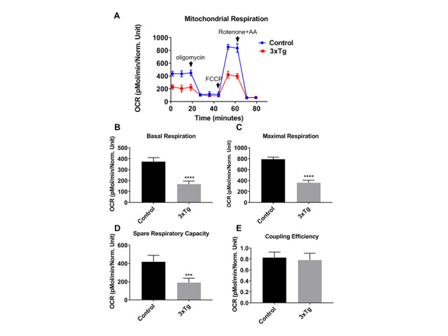
Current treatments targeting amyloid beta in Alzheimer's disease (AD) have minimal efficacy, which results in a huge unmet medical need worldwide. Accumulating data suggest that brain mitochondrial dysfunction play a critical role in AD pathogenesis. Targeting cellular mechanisms associated with mitochondrial dysfunction in AD create a novel approach for drug development. This study investigated the effects of nilotinib, as a selective tyrosine kinase inhibitor, in astroglia derived from 3xTg-AD mice versus their C57BL/6-controls. Parameters included oxygen consumption rates (OCR), ATP, cytochrome c oxidase (COX), citrate synthase (CS) activity, alterations in oxidative phosphorylation (OXPHOS), nuclear factor kappa B (NF-κB), key regulators of mitochondrial dynamics (mitofusin (Mfn1), dynamin-related protein 1 (Drp1)), and mitochondrial biogenesis (peroxisome proliferator-activated receptor gamma coactivator1-alpha (PGC-1α), calcium/calmodulin-dependent protein kinase II (CaMKII), and nuclear factor (erythroid-derived 2)-like 2 (Nrf2)). Nilotinib increased OCR, ATP, COX, Mfn1, and OXPHOS levels in 3xTg astroglia. No significant differences were detected in levels of Drp1 protein and CS activity. Nilotinib enhanced mitochondrial numbers, potentially through a CaMKII-PGC1α-Nrf2 pathway in 3xTg astroglia. Additionally, nilotinib-induced OCR increases were reduced in the presence of the NF-κB inhibitor, Bay11-7082. The data suggest that NF-κB signaling is intimately involved in nilotinib-induced changes in bioenergetics in 3xTg brain astroglia. Nilotinib increased translocation of the NF-κB p50 subunit into the nucleus of 3xTg astroglia that correlates with an increased expression and activation of NF-κB. The current findings support a role for nilotinib in improving mitochondrial function and suggest that astroglia may be a key therapeutic target in treating AD.

Keywords: Alzheimer’s disease; astroglia; bioenergetics; biogenesis; citrate synthase; cytochrome c oxidase; mitochondrial function; nuclear factor kappa B (NF-κB); oxidative phosphorylation.


Conflict of interest statement

Conflict of Interest The authors declare no conflict of interest.

Figures

Figure 1.
Figure 1.
Mitochondrial respiration rates are reduced in 3xTg-AD astroglia. (A) Kinetics graph indicating real-time OCR at baseline and after addition of oligomycin, FCCP, and rotenone/antimycin. The OCR was measured in 3xTg and C57BL/6-WT astroglia utilizing the XF24 analyzer. (B) Basal respiration, (C) maximal respiration, (D) spare respiratory capacity, and (E) Coupling efficiency were calculated compared between 3xTg and control cells. Data are mean ± SD of n = 6 per group (*** = p < 0.001 or **** = p < 0.0001) analyzed by unpaired Student's t-test.
Figure 2.
Figure 2.
Nilotinib did not alter mitochondrial respiration rate or expression of total or phospho c-Abl in C57BL/6-WT astroglia. The OCR was measured in C57BL/6 astroglia utilizing the XF24 analyzer after 24 hours treatment with nilotinib (1, 10, 100, and 1000 nM). (A) Basal respiration, (B) maximal respiration, (C) spare respiratory capacity, (D) Coupling efficiency, and (E) total ATP level were calculated and compared between control and treated cells. Western blot experiments demonstrating relative levels of c-Abl and p-c-Abl in C57BL/6-WT astroglia (F) in the presence and absence of 24 hrs. 100 nM nilotinib treatment. Relative quantification for protein levels of c-Abl and p-c-Abl normalized to total protein and total c-Abl, respectively (G, H). Data are mean ± SD of n = 6 per group (* = p < 0.05) analyzed by unpaired Student's t-test.
Figure 3.
Figure 3.
Nilotinib improved mitochondrial respiration rates in 3xTg-AD astroglia. Nilotinib inhibited the expression level of phospho c-Abl in 3xTg astroglia. The OCR was measured in 3xTg-AD astroglia utilizing the XF24 analyzer after 24 hours treatment with nilotinib (1, 10, 100, and 1000 nM). (A) Basal respiration, (B) maximal respiration, (C) spare respiratory capacity, (D) coupling efficiency, and (E) total ATP level were calculated and compared between control and treated cells. Western blot experiments demonstrating relative levels of c-Abl and p-c-Abl in 3xTg-AD astroglia (F) in the presence and absence of 24 hrs. 100 nM nilotinib treatment. Relative quantification for protein levels of c-Abl and p-c-Abl normalized to total protein and total c-Abl, respectively (G, H). Data are mean ± SD of n = 6 per group (* = p < 0.05 or ** = p < 0.01 or *** = p < 0.001 or **** = p < 0.0001) analyzed by unpaired Student's t-test.
Figure 4.
Figure 4.
Nilotinib did not alter the expression of mitochondrial Complex (I-V) protein subunits in C57BL/6-WT astroglia. Western blot experiments demonstrating relative levels of mitochondrial protein subunits in C57BL/6-WT astroglia in the presence and absence of 24 hrs. 100 nM nilotinib treatment. (A) Representative Western blot for NADH dehydrogenase beta sub complex subunit 8 of Complex I (NDUFB8), succinate dehydrogenase subunit B of Complex II (SDHB), cytochrome b-c1 complex subunit 2 of Complex III (UQCRC2), Cytochrome c oxidase subunit 1 of Complex IV (MTCO1), and ATP synthase subunit alpha of Complex V (ATP5A). (B-F) Relative quantification for protein levels of Complex I-V normalized to total protein. (G) COX activity was measured in C57BL/6-WT astroglia in presence and absence of 100 nM nilotinib treatment. Results are expressed as mean ± SD of n = 6 per group (*P ≤ 0.05) analyzed by unpaired Student's t-test.
Figure 5.
Figure 5.
Nilotinib significantly increased the expression of mitochondrial Complex (I and III-V) protein subunits and cytochrome c oxidase (COX) activity in 3xTg-AD astroglia. Western blot experiments demonstrating relative levels of mitochondrial protein subunits in 3xTg astroglia in the presence and absence of 24 hrs. 100 nM nilotinib treatment. (A) Representative Western blot for NADH dehydrogenase beta subcomplex subunit 8 of Complex I (NDUFB8), succinate dehydrogenase subunit B of Complex II (SDHB), cytochrome b-c1 complex subunit 2 of Complex III (UQCRC2), Cytochrome c oxidase subunit 1 of Complex IV (MTCO1), and ATP synthase subunit alpha of Complex V (ATP5A). (B-F) Relative quantification for protein levels of Complex I-V normalized to total protein. (G) COX activity was measured in 3xTg-AD astroglia in presence and absence of 100 nM nilotinib treatment. Results are expressed as mean ± SD of n = 6 per group (*P ≤ 0.05**P ≤ 0.01) analyzed by unpaired Student's t-test.
Figure 6.
Figure 6.
Nilotinib significantly altered the expression of Mfn1 in 3xTg-AD astroglia. Western blot experiments demonstrating relative levels of Mfn1 and Drp1 in C57BL/6-WT astroglia (A-C) and 3xTg astroglia (D-F) inthe presence and absence of 24 hrs. 100 nM nilotinib treatment. Relative quantification for protein levels of Mfn-1 and Drp-1 normalized to total protein. Results are expressed as mean ± SD of n = 6 per group (***P ≤ 0.001) analyzed by unpaired Student's t-test.
Figure 7.
Figure 7.
Nilotinib increased the number of mitochondria and mitochondrial biogenesis (CAMKII, PGC1-α, and Nrf2) in 3xTg-AD astroglia. The numbers of mitochondria in 3xTg astroglia were compared in the presence and absence of nilotinib (100 nM). Representative TEM image A showing the cell body of 3xTg astroglia. Representative TEM image B showing the cell body of the nilotinib-treated 3xTg-AD astroglia. (C) The number of mitochondria per cross-sectioned cell was counted (20 cells per group counted by TEM). Results are expressed as mean ± SD of n=20 per group (****P ≤ 0.0001) analyzed by unpaired Student's t-test. M: mitochondria; Scale bars: 500 μm; Magnification: 46000x. (D-H) Western blot experiments demonstrating relative levels of CAMKII, PGC1-α, TFAM, and Nrf2 in 3xTg-AD astroglia in the presence and absence of 24 hrs. 100 nM nilotinib treatment. Relative quantification for protein levels of CAMKII, PGC1-α, TFAM, and Nrf2 normalized to total protein. Results are expressed as mean ± SD of n = 5 per group (* = p < 0.05 or ** = p < 0.01 or **** = p < 0.0001) analyzed by unpaired Student's t-test.
Figure 8.
Figure 8.
Nilotinib significantly increased the activation of NF-κB and expression of NF-κB p50/p105 subunits in C57BL/6-WT astroglia. (A) Western blot experiments demonstrating relative levels of NF-κB subunits (p50, p105, p65, and p75), and IκB-α in cultured cortical astroglia derived from C57BL/6 in the presence and absence of 24 hrs. 100 nM nilotinib treatment. (B-F) Relative quantification for protein levels of NF-κB subunits (p50, p105, p65, and p75) and IκB-α normalized to total protein. (G) Nuclear extract derived from nilotinib-treated and non-treated C57BL/6-WT astroglia were assayed for NF-κB activation by EMSA using a biotin-labeled oligonucleotide encompassing the NF-κB consensus motif. Results are expressed as mean ± SD of n = 6 per group (**P ≤ 0.01) analyzed by unpaired Student's t-test.
Figure 9.
Figure 9.
Nilotinib significantly increased the expression of NF-κB p50/p105 subunits and activation of NF-κB in 3xTg-AD astroglia. (A) Western blot experiments demonstrating relative levels of NF-κB subunits (p50, p105, p65, and p75), and IκB-α in cultured cortical astroglia derived from 3xTg in the presence and absence of 24 hrs.100 nM nilotinib treatment. (B-F) Relative quantification for protein levels of NF-κB subunits (p50, p105, p65, and p75) and IκB-α normalized to total protein. (G) Nuclear extract derived from nilotinibtreated and non-treated 3xTg-AD astroglia were assayed for NF-κB activation by EMSA using a biotinlabeled oligonucleotide encompassing the NF-κB consensus motif. Results are expressed as mean ± SD of n = 6 per group (*P ≤ 0.05) analyzed by unpaired Student's t-test.
Figure 10.
Figure 10.
Nilotinib translocated the NF-kB p50 subunit into the nucleus of 3xTg-AD astroglia. Quantitative immunofluorescent nuclear/cytoplasmic ratios of NF-κB subunits (p50 (A), p65 (B), and p75 (C)) in 3xTg-AD astroglia. DAPI (blue) marks the nucleus. Images were captured at 100x magnification. Volume density of NF-kB subunits immunofluorescence was quantified using ImageJ software (****P ≤ 0.0001); n=5 per group analyzed by unpaired Student's t-test.
Figure 11.
Figure 11.
Bay11-7082 decreased mitochondrial respiration rates in 3xT-AD astroglia. (A) Kinetics graph indicating real-time OCR at baseline and after addition of oligomycin, FCCP, and rotenone/antimycin. The OCR was measured in 3xTg-AD astroglia utilizing the XF24 analyzer after 24 hours treatment with Bay11- 7082 (1, 3, 5, and 10 uM). (B) Basal respiration, (C) maximal respiration and (D) spare respiratory capacity, and (E) coupling efficiency were calculated compared between control and treated cells. Data are mean ± SD of n = 5 group (** = p < 0.01) analyzed by unpaired Student's t-test.
Figure 12.
Figure 12.
Nilotinib-induced enhancements in energy metabolism appear NF-kB-dependent, as demonstrated in 3xTg-AD astroglia. The OCR was measured in 3xTg-AD astroglia utilizing the XF24 analyzer after 24 hours treatment with nilotinib (100 nM) or Bay11-7082 (5 uM) or co-treatment with nilotinib (100 nM) + Bay11- 7082 (5 uM). (A) Basal respiration, (B) maximal respiration, (C) spare respiratory capacity, (D) coupling efficiency, and (E) ATP level were calculated compared between control and treated cells. Data are mean ± SD of n= 5 per group (* = p < 0.05 or ** = p < 0.01 or *** = p < 0.001 or **** = p < 0.0001) analyzed by unpaired Student's t-test.
Figure 13.
Figure 13.
This putative pathway suggests that nilotinib improves mitochondrial function through nuclear factor κ B (NF-κB)-dependent signaling in 3xTg-AD astroglia. It is hypothesized that nilotinib activates CAMKII as well as a signal cascade resulting in degradation of the NF-κB subunit IκB. The NF-κB dimer then translocates from the cytoplasm to the nucleus where it binds to a DNA consensus sequence of target genes. It is also hypothesized that nilotinib triggers the NF-κB complex (downstream target of CAMKII) to move into the mitochondrion, where is thought to interact with OXPHOS genes that leads to bioenergetic improvement (ATP, oxygen consumption rate (OCR)), as well as an increase in the expression of mitochondrial complex proteins subunits (Complexes I, III, IV, and V), cytochrome c oxidase (COX), mitochondrial biogenesis (PGC1-α and Nrf2), and mitochondrial dynamics (Mfn1). Findings from our current study importantly show that mitochondrial OCR and ATP levels were significantly reduced in the presence of the IKK inhibitor (Bay11-7082). This figure was developed using the BioRender online software tool.

Similar articles

Cited by

References

    1. McKeith I, Cummings J (2005). Behavioural changes and psychological symptoms in dementia disorders. Lancet Neurol, 4:735-742. - PubMed
    1. Glabe CC (2005). Amyloid accumulation and pathogensis of Alzheimer's disease: significance of monomeric, oligomeric and fibrillar Abeta. Subcell Biochem, 38:167-177. - PubMed
    1. Hardy J, Selkoe DJ (2002). The amyloid hypothesis of Alzheimer's disease: progress and problems on the road to therapeutics. Science, 297:353-356. - PubMed
    1. Cardoso S, Seica RM, Moreira PI (2017). Mitochondria as a target for neuroprotection: implications for Alzheimer s disease. Expert Rev Neurother, 17:77-91. - PubMed
    1. Cadonic C, Sabbir MG, Albensi BC (2016). Mechanisms of Mitochondrial Dysfunction in Alzheimer's Disease. Mol Neurobiol, 53:6078-6090. - PubMed

LinkOut - more resources