

Advantages of preoperative planning using computed tomography scan for treatment of malleolar ankle fractures
- PMID: 33816140
- PMCID: PMC7995337
- DOI: 10.5312/wjo.v12.i3.129
Advantages of preoperative planning using computed tomography scan for treatment of malleolar ankle fractures
Abstract
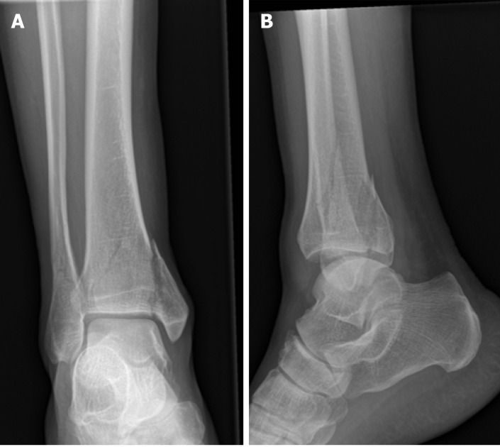
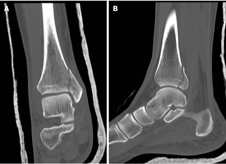
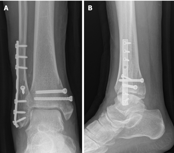
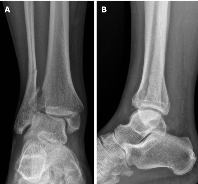
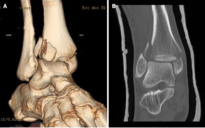
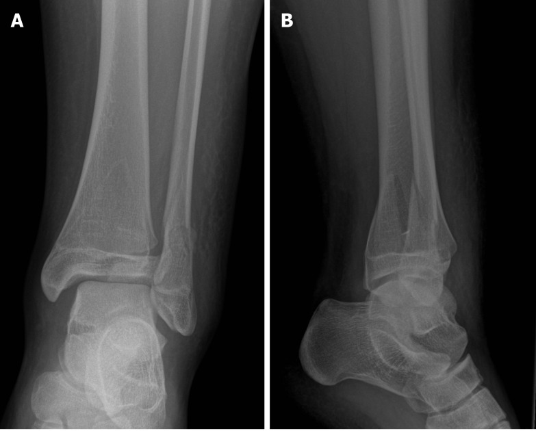
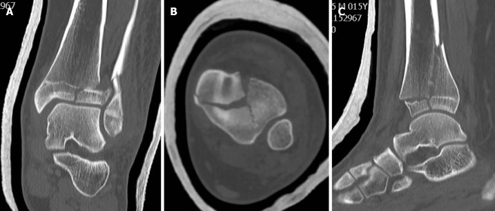
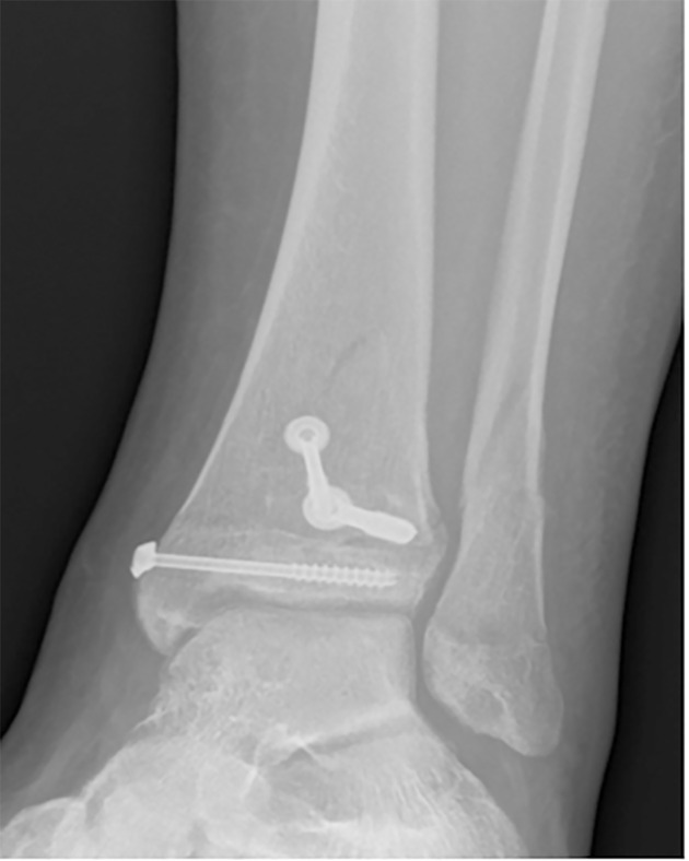
Malleolar ankle fractures have been classified using plain radiographs, and there is no consensus regarding the role of computed tomography (CT) scans in preoperative planning. We analyzed critical aspects, such as limits of standard radiographs, types of injury, classification methods and cost/benefit evaluations. CT scans allow a 3D analysis of the fracture to be obtained and consequently assess the indication for surgical procedure, surgical access and the type of fixation devices required. This exam is useful for detecting lesions that may go unnoticed on radiographs and will help surgeons to clarify the pathoanatomy of ankle fractures. According to Arbeitsgemeinschaft fur Osteosynthesefragen/ Orthopaedic Trauma Association (AO/OTA) classification, CT scan is recommended in medial malleolar fractures with vertical rim, type 44B fractures with posterior malleolar involvement and all type 44C fractures (according to AO/OTA). Also Tillaux-Chaput fractures (43-B1 according to AO/OTA), malleolar fractures in the presence of distal tibial fractures (43 according to AO/OTA) and distal tibia fractures in adolescents should be studied with CT scans.
Keywords: Computed tomography scan; Imaging; Malleolar fractures; Planning; Trauma.
©The Author(s) 2021. Published by Baishideng Publishing Group Inc. All rights reserved.
Conflict of interest statement
Conflict-of-interest statement: The authors have no conflicts of interest to declare.
Figures
References
-
- Elsoe R, Ceccotti AA, Larsen P. Population-based epidemiology and incidence of distal femur fractures. Int Orthop. 2018;42:191–196. - PubMed
-
- Black EM, Antoci V, Lee JT, Weaver MJ, Johnson AH, Susarla SM, Kwon JY. Role of preoperative computed tomography scans in operative planning for malleolar ankle fractures. Foot Ankle Int. 2013;34:697–704. - PubMed
-
- Court-Brown CM, Caesar B. Epidemiology of adult fractures: A review. Injury. 2006;37:691–697. - PubMed
-
- Jensen SL, Andresen BK, Mencke S, Nielsen PT. Epidemiology of ankle fractures. A prospective population-based study of 212 cases in Aalborg, Denmark. Acta Orthop Scand. 1998;69:48–50. - PubMed
-
- Driesman AS, Egol KA. An update on the treatment of malleolar fractures. Fuss Sprungg. 2016;14:55–65.
Publication types
LinkOut - more resources
Full Text Sources
Other Literature Sources
