

Dual-energy computed tomography of the neck-optimizing tube current settings and radiation dose using a 3D-printed patient phantom
- PMID: 33816156
- PMCID: PMC7930688
- DOI: 10.21037/qims-20-854
Dual-energy computed tomography of the neck-optimizing tube current settings and radiation dose using a 3D-printed patient phantom
Abstract
Background: Dual-energy computed tomography (DECT) is increasingly used in studies and clinical practice. However, the best protocol is controversially discussed and whether it exhibits more radiation exposure compared to conventional protocols. Thus, the purpose of the study was to determine optimal tube current settings for DECT in a 3D-printed anthropomorphic phantom of the neck.
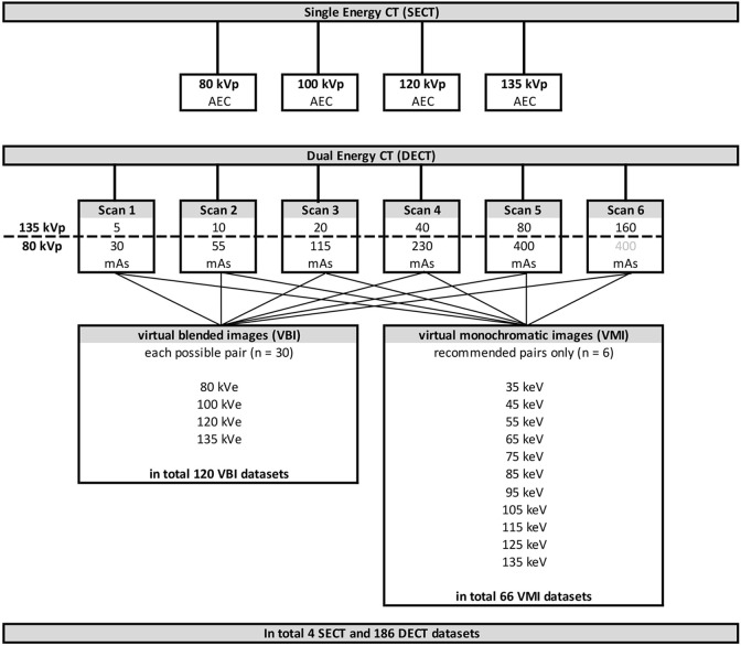
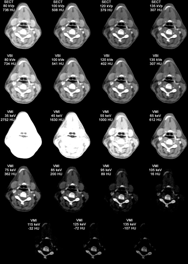
Methods: A 3D-printed iodinated ink based phantom of a contrast enhanced CT of the neck was imaged. Six dual-energy multi-detector computed tomography scans were performed with six different tube currents (80 kVp: 30-400 mAs; 135 kVp: 5-160 mAs). 120 virtual blended images (VBIs) and 66 virtual monochromatic images (VMIs) were reconstructed and 12 regions of interest (bilaterally: common carotid arteries, subcutaneous soft tissue, mandibular bone, sternocleidomastoid muscle, submandibular gland, and mid-image: vertebral body of C2 and pharyngeal space) in six consecutive slices resulting in 96 measurements per scan were performed. Hounsfield units and signal- and contrast-to-noise ratio were compared to single-energy computed tomography as standard of reference.
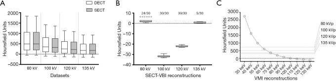
Results: VBIs overestimated the Hounsfield units (P<0.0001). Optimal dual-energy scanning parameters resulted in 120% (100 kVe: 51.2 vs. 61.7 and 65.2, for signal and contrast-to-noise ratio, respectively; 120 kVe: 60.8 vs. 72.1 vs. 128.3) of the radiation exposure with about 80% of the signal/contrast-to-noise ratio of the corresponding single-energy images. However, optimal weighting of tube currents for both voltages depended on the desired reconstruction.
Conclusions: Dual-energy protocols apply an estimated 120% of the single-energy radiation exposure and result in approximately 80% of the image quality. Tube current settings should be adapted to the desired information.
Keywords: Phantoms; X-ray computed; imaging; printing; radiation exposure; three-dimensional; tomography.
2021 Quantitative Imaging in Medicine and Surgery. All rights reserved.
Conflict of interest statement
Conflicts of Interest: All authors have completed the ICMJE uniform disclosure form (available at http://dx.doi.org/10.21037/qims-20-854). TD held talks for Canon Medical System. However, the company did not influence nor authorize the results of this study. MS reports grants from Bundesministerium für Wirtschaft und Energie (DE), during the conduct of the study; other from PhantomX GmbH, outside the submitted work; in addition, MS has a patent DE202015104282U1 issued, a patent US9924919B2 issued, a patent US10182786B2 issued, and a patent EP3135199A1 pending. BH reports grants to the Department of Radiology outside the submitted work from Abbott, Actelion Pharma, Bayer Schering Pharma, Bayer Vital, BRACCO Group, Bristol-Myers Squibb, Charité Research organisation GmbH, Deutsche Krebshilfe, Dt. Stiftung für Herzforschung, Essex Pharma, EU Programmes, Fibrex Medical Inc., Focused ultrasound Surgery Foundation, Fraunhofer Gesellschaft, Gurbet, INC Research, InSightec Ltd., IPSEN Pharma, Kendle/MorphoSys AG, Lilly GmbH, Lundbeck GmbH, MeVis Medical Solutions AG, Nexus Oncology, Novartis, Parexel CRO Service, Perceptive, Pfizer GmbH, Philipps, sonofis-aventis S.A., Siemens, Spectranetics GmbH, Terumo Medical Corporation, TNS Healthcare GmbH, Canon Medical, UCB Pharma, Wyeth Pharma, Zukunftsfond Berlin (TSB), outside the submitted work. Dr. PJ reports grants from Bundesministerium für Wirtschaft und Energie (DE), during the conduct of the study; other from PhantomX GmbH, outside the submitted work; in addition, PJ has a patent DE202015104282U1 issued, a patent US9924919B2 issued, a patent US10182786B2 issued, and a patent EP3135199A1 pending. The other author has no conflicts of interest to declare.
Figures


Similar articles
-
Image quality comparison between single energy and dual energy CT protocols for hepatic imaging.Med Phys. 2016 Aug;43(8):4877. doi: 10.1118/1.4959554. Med Phys. 2016. PMID: 27487905
-
Quantitative accuracy and dose efficiency of dual-contrast imaging using dual-energy CT: a phantom study.Med Phys. 2020 Feb;47(2):441-456. doi: 10.1002/mp.13912. Epub 2019 Dec 10. Med Phys. 2020. PMID: 31705664 Free PMC article.
-
Virtual monochromatic imaging in dual-source dual-energy CT: radiation dose and image quality.Med Phys. 2011 Dec;38(12):6371-9. doi: 10.1118/1.3658568. Med Phys. 2011. PMID: 22149820 Free PMC article.
-
Can virtual monochromatic images from dual-energy CT replace low-kVp images for abdominal contrast-enhanced CT in small- and medium-sized patients?Eur Radiol. 2019 Jun;29(6):2878-2889. doi: 10.1007/s00330-018-5850-z. Epub 2018 Nov 30. Eur Radiol. 2019. PMID: 30506223
-
Determination of the optimal range for virtual monoenergetic images in dual-energy CT based on physical quality parameters.Med Phys. 2021 Sep;48(9):5085-5095. doi: 10.1002/mp.15120. Epub 2021 Aug 4. Med Phys. 2021. PMID: 34287956
Cited by
-
Interdisciplinary challenges and aims of flap or graft reconstruction surgery of sinonasal cancers: What radiologists and radiation oncologists need to know.Front Oncol. 2022 Sep 20;12:1013801. doi: 10.3389/fonc.2022.1013801. eCollection 2022. Front Oncol. 2022. PMID: 36203460 Free PMC article. Review.
-
Optimization of Fast Non-Local Means Noise Reduction Algorithm Parameter in Computed Tomographic Phantom Images Using 3D Printing Technology.Diagnostics (Basel). 2024 Jul 23;14(15):1589. doi: 10.3390/diagnostics14151589. Diagnostics (Basel). 2024. PMID: 39125465 Free PMC article.
References
LinkOut - more resources
Full Text Sources
Other Literature Sources
Miscellaneous