Meynert nucleus-related cortical thinning in Parkinson's disease with mild cognitive impairment
- PMID: 33816191
- PMCID: PMC7930701
- DOI: 10.21037/qims-20-444
Meynert nucleus-related cortical thinning in Parkinson's disease with mild cognitive impairment
Abstract
Background: Cognitive impairment in Parkinson's disease (PD) involves the cholinergic system and cholinergic neurons, especially the nucleus basalis of Meynert (NBM/Ch4) located in the basal forebrain (BF). We analyzed associations between NBM/Ch4 volume and cortical thickness to determine whether the NBM/Ch4-innervated neocortex shows parallel atrophy with the NBM/Ch4 as disease progresses in PD patients with cognitive impairment (PD-MCI).
Methods: We enrolled 35 PD-MCI patients, 48 PD patients with normal cognition (PD-NC), and 33 age- and education-matched healthy controls (HCs), with all participants undergoing neuropsychological assessment and structural magnetic resonance imaging (MRI). Correlation analyses between NBM/Ch4 volume and cortical thickness and correlation coefficient comparisons were conducted within and across groups.
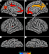
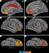
Results: In the PD-MCI group, NBM/Ch4 volume was positively correlated with cortical thickness in the bilateral posterior cingulate, parietal, and frontal and left insular regions. Based on correlation coefficient comparisons, the atrophy of NBM/Ch4 was more correlated with the cortical thickness of right posterior cingulate and precuneus, anterior cingulate and medial orbitofrontal lobe in PD-MCI versus HC, and the right medial orbitofrontal lobe and anterior cingulate in PD-NC versus HC. Further partial correlations between cortical thickness and NBM/Ch4 volume were significant in the right medial orbitofrontal (PD-NC: r=0.3, P=0.045; PD-MCI: r=0.51, P=0.003) and anterior cingulate (PD-NC: r=0.41, P=0.006; PD-MCI: r=0.43, P=0.013) in the PD groups and in the right precuneus (r=0.37, P=0.04) and posterior cingulate (r=0.46, P=0.008) in the PD-MCI group.
Conclusions: The stronger correlation between NBM/Ch4 and cortical thinning in PD-MCI patients suggests that NBM/Ch4 volume loss may play an important role in PD cognitive impairment.
Keywords: Parkinson’s disease (PD); cholinergic system; cognitive impairment; cortical thickness; nucleus basalis of Meynert.
2021 Quantitative Imaging in Medicine and Surgery. All rights reserved.
Conflict of interest statement
Conflicts of Interest: All authors have completed the ICMJE uniform disclosure form (available at http://dx.doi.org/10.21037/qims-20-444). The authors have no conflicts of interest to declare.
Figures


References
-
- Lawson RA, Yarnall AJ, Duncan GW, Breen DP, Khoo TK, Williams-Gray CH, Barker RA, Collerton D, Taylor JP, Burn DJ, ICICLE-PD study group . Cognitive decline and quality of life in incident Parkinson's disease: The role of attention. Parkinsonism Relat Disord 2016;27:47-53. 10.1016/j.parkreldis.2016.04.009 - DOI - PMC - PubMed
LinkOut - more resources
Full Text Sources
Other Literature Sources
Miscellaneous
