. 2021 Mar 19:11:616156.
doi: 10.3389/fonc.2021.616156. eCollection 2021.

MR-Guided Radiotherapy for Head and Neck Cancer: Current Developments, Perspectives, and Challenges

Affiliations

MR-Guided Radiotherapy for Head and Neck Cancer: Current Developments, Perspectives, and Challenges

Simon Boeke et al. Front Oncol. .

Abstract

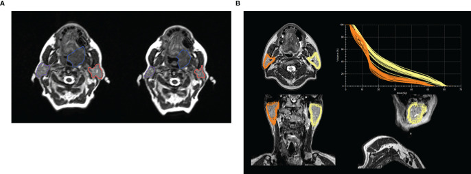
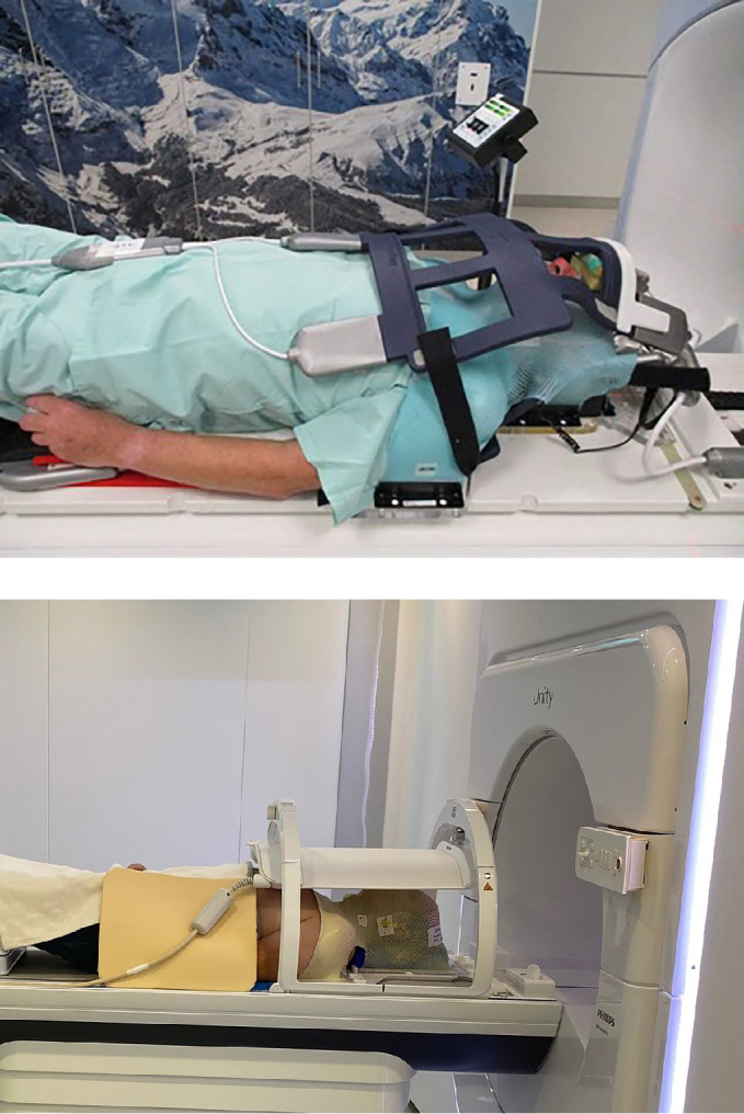
Based on the development of new hybrid machines consisting of an MRI and a linear accelerator, magnetic resonance image guided radiotherapy (MRgRT) has revolutionized the field of adaptive treatment in recent years. Although an increasing number of studies have been published, investigating technical and clinical aspects of this technique for various indications, utilizations of MRgRT for adaptive treatment of head and neck cancer (HNC) remains in its infancy. Yet, the possible benefits of this novel technology for HNC patients, allowing for better soft-tissue delineation, intra- and interfractional treatment monitoring and more frequent plan adaptations appear more than obvious. At the same time, new technical, clinical, and logistic challenges emerge. The purpose of this article is to summarize and discuss the rationale, recent developments, and future perspectives of this promising radiotherapy modality for treating HNC.

Keywords: IGRT (Image Guided Radiation Therapy); MR-guidance; MRI; adaptive radiotherapy; head and neck (H&N) cancer; salivary gland; xerostoma.


Conflict of interest statement

The authors declare that the Department of Radiation Oncology Tübingen receives within the frame of research agreements financial and technical support as well as sponsoring for travels and scientific symposia from Elekta AB (Stockholm, Sweden), TheraPanacea (Paris, France), Philips GmbH (Best, The Netherlands), Dr. Sennewald Medizintechnik GmbH (München, Germany), PTW Freiburg (Germany) and that the Department of Radiation Oncology of the Zurich University Hospital receives within the frame of research agreements funding for clinical trials from ViewRay Inc, Oakwood, USA. PB received research funding from ViewRay Inc, Oakwood, USA.

Figures

Figure 1
Figure 1
(A) Example of volume changes and migration of parotid glands during the course of fractionated radiotherapy at an 0.35 T MR-Linac or a large base of tongue carcinoma between treatment start (left image) and beginning of the 7th treatment week - boost (right image). Left and right parotid glands are delineated in orange and violet respectively and the gross tumor volume in blue. The volume of the left and right parotid glands decreased by 8.2 cc and 10.0 cc, respectively. The inter-parotids distance changed from 11.0 cm to 10.3 cm. (B) Example of a post treatment analysis for a patient treated for a hypopharyngeal carcinoma with 70 Gy in 35 fractions. Parotid glands were contoured for each daily MRI during the course of fractionated radiotherapy at a 1.5 T MR-Linac and propagated to the T2w planning MRI, with the total plan DVH for each daily delivered plan in the upper right corner, showing the variance in actual delivered dose depending on volume of the parotid gland. Averaged Dmean of the anatomically corrected and daily adapted plans was 24.4 Gy and 16.5 Gy for the left and right parotid glands, respectively. The Dmean of the reference plan was 25.9 Gy for the left and 16.7 Gy for the right parotid gland. Baseline volume was 31.0 ccm for the right and 34.5 ccm for the left parotid gland. Mean volume (range) during treatment was 30.3 ccm (29.5–32.1) and 31.4 ccm (29.1–34.7). The example was presented as a poster at the congresses of DEGRO and AIRO 2019 by Monica lo Russo, MD (20).
Figure 2
Figure 2
Patient positioning for MR-Linac based treatment for head and neck cancer in the two commercially available systems.

References

    1. Grégoire V, Guckenberger M, Haustermans K, Lagendijk JJW, Ménard C, Pötter R, et al. . Image guidance in radiation therapy for better cure of cancer. Mol Oncol (2020) 14:1470–91. 10.1002/1878-0261.12751 - DOI - PMC - PubMed
    1. Chin S, Eccles CL, McWilliam A, Chuter R, Walker E, Whitehurst P, et al. . Magnetic resonance-guided radiation therapy: A review. J Med Imaging Radiat Oncol (2020) 64:163–77. 10.1111/1754-9485.12968 - DOI - PubMed
    1. Mutic S, Dempsey JF. The ViewRay system: magnetic resonance-guided and controlled radiotherapy. Semin Radiat Oncol (2014) 24:196–9. 10.1016/j.semradonc.2014.02.008 - DOI - PubMed
    1. Raaymakers BW, Jürgenliemk-Schulz IM, Bol GH, Glitzner M, Kotte ANTJ, van Asselen B, et al. . First patients treated with a 1.5 T MRI-Linac: clinical proof of concept of a high-precision, high-field MRI guided radiotherapy treatment. Phys Med Biol (2017) 62:L41–50. 10.1088/1361-6560/aa9517 - DOI - PubMed
    1. Corradini S, Alongi F, Andratschke N, Belka C, Boldrini L, Cellini F, et al. . MR-guidance in clinical reality: current treatment challenges and future perspectives. Radiat Oncol Lond Engl (2019) 14:92. 10.1186/s13014-019-1308-y - DOI - PMC - PubMed

LinkOut - more resources