

Bladder wall micromotion measured by non-invasive ultrasound: initial results in women with and without overactive bladder
- PMID: 33816693
- PMCID: PMC8012835
Bladder wall micromotion measured by non-invasive ultrasound: initial results in women with and without overactive bladder
Abstract
Objective: Rhythmic contractions of the bladder wall during filling result from the synchronization of bladder wall micromotion and are often observed in the urodynamic tracings of individuals with urinary overactive bladder (OAB). This study's objective was to develop a novel, non-invasive method to measure bladder wall micromotion and to conduct an initial study to test the hypothesis that elevated micromotion is associated with OAB.
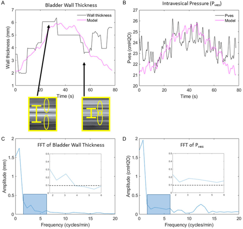
Methods: This prospective study enrolled women with OAB and asymptomatic volunteers as measured by the ICIQ-OAB survey. After filling the bladder to 40% cystometric capacity, 85 second cine-loops were obtained using a GE Voluson E8 ultrasound system with an 8 MHz curved, abdominal probe. A custom correlation-based texture tracking MATLAB algorithm was used to measure changes in the bladder wall thickness over time and correlate with changes in vesical pressure. Significant bladder wall micromotion was defined as changes in wall thickness with amplitudes higher than 0.1 mm in the frequency range of 1.75-6 cycles/minute as calculated from Fast Fourier Transform (FFT) analysis. The micromotion algorithm was tested on 30 women including 17 with OAB and 13 asymptomatic volunteers.
Results: Micromotion was identified in 41% of subjects with OAB and 0% of asymptomatic volunteers, indicating a significant association of micromotion with OAB (Fisher's exact test, P=0.010). Micromotion was also found to have a significant association with a clinical diagnosis of detrusor overactivity (Fisher's exact test, P=0.031). Frequencies with elevated micromotion correlated with frequencies of vesical pressure fluctuations.
Conclusions: The feasibility of a non-invasive method to measure bladder wall micromotion was demonstrated using transabdominal anatomical motion mode (M-mode) ultrasound. Presence of micromotion was significantly associated with OAB and with urodynamic-identified rhythm.
Keywords: Ultrasonography; detrusor overactivity; diagnostic imaging; fast Fourier transform analysis; image analysis; lower urinary tract symptoms; non-invasive; overactive bladder; texture tracking; urodynamics.
AJCEU Copyright © 2021.
Conflict of interest statement
None.
Figures


Comment in
-
Voiding Function and Dysfunction, Bladder Physiology and Pharmacology, and Female Urology.J Urol. 2021 Oct;206(4):1067-1070. doi: 10.1097/JU.0000000000002134. Epub 2021 Jul 21. J Urol. 2021. PMID: 34284648 No abstract available.
References
-
- Coyne KS, Sexton CC, Vats V, Thompson C, Kopp ZS, Milsom I. National community prevalence of overactive bladder in the United States stratified by sex and age. Urology. 2011;77:1081–1087. - PubMed
-
- Abrams P, Kelleher CJ, Kerr LA, Rogers RG. Overactive bladder significantly affects quality of life. Am J Manag Care. 2000;6(Suppl):S580–590. - PubMed
-
- Schabert VF, Bavendam T, Goldberg EL, Trocio JN, Brubaker L. Challenges for managing overactive bladder and guidance for patient support. Am J Manag Care. 2009;15(Suppl):S118–122. - PubMed
-
- Abrams P. Describing bladder storage function: overactive bladder syndrome and detrusor overactivity. Urology. 2003;62:28–37. discussion 40-22. - PubMed
-
- Hashim H, Abrams P. Is the bladder a reliable witness for predicting detrusor overactivity? J Urol. 2006;175:191–194. discussion 194-195. - PubMed
Grants and funding
LinkOut - more resources
Full Text Sources