

The Treatment of Muscle Atrophy after Rotator Cuff Tears Using Electroconductive Nanofibrous Matrices
- PMID: 33816776
- PMCID: PMC8011566
- DOI: 10.1007/s40883-020-00186-8
The Treatment of Muscle Atrophy after Rotator Cuff Tears Using Electroconductive Nanofibrous Matrices
Abstract
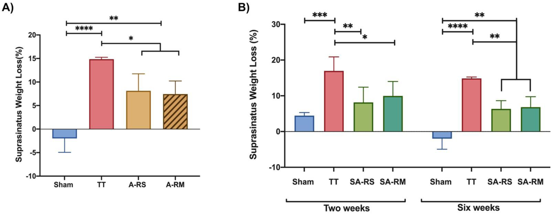
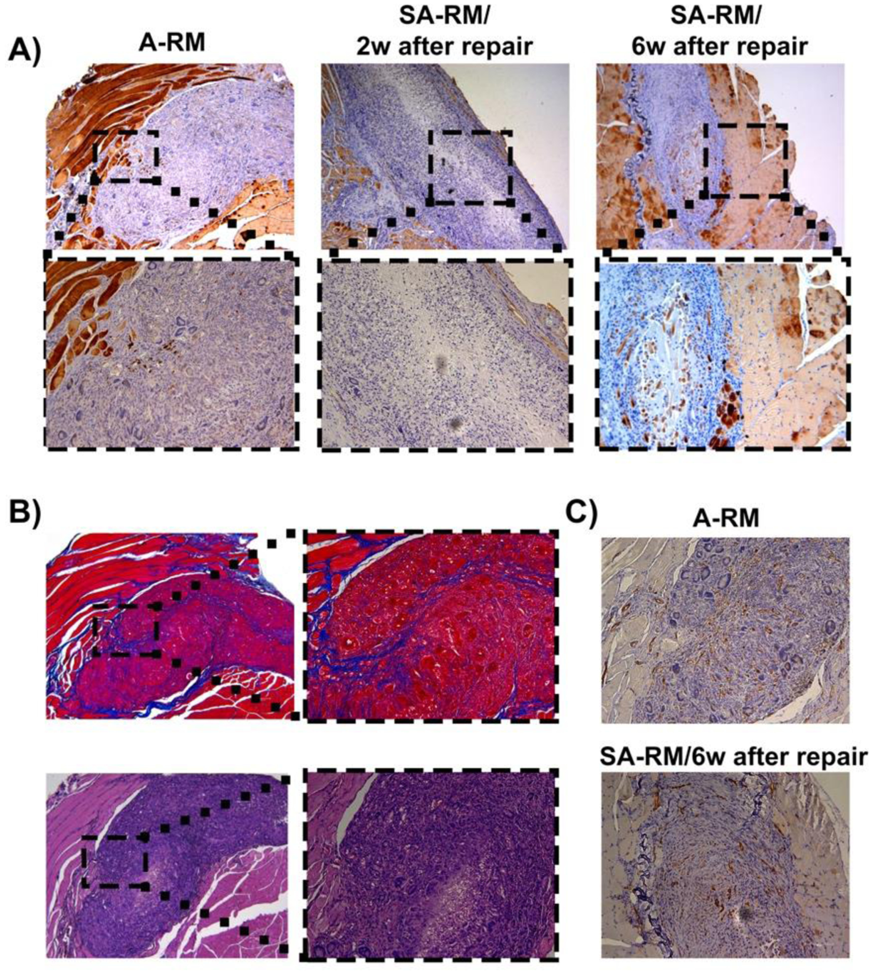
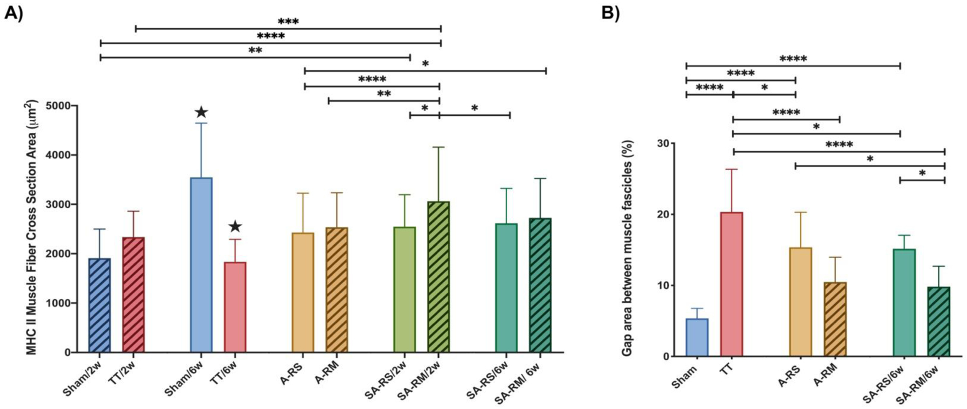
Rotator cuff tears (RCTs) are a common cause of disability and pain in the adult population. Despite the successful repair of the torn tendon, the delay between the time of injury and time of repair can cause muscle atrophy. The goal of the study was to engineer an electroconductive nanofibrous matrix with an aligned orientation to enhance muscle regeneration after rotator cuff (RC) repair. The electroconductive nanofibrous matrix was fabricated by coating Poly(3,4-ethylenedioxythiophene): poly(styrenesulfonate) (PEDOT:PSS) nanoparticles onto the aligned poly(ε-caprolactone) (PCL) electrospun nanofibers. The regenerative potential of the matrix was evaluated using two repair models of RCTs include acute and sub-acute. Sprague-Dawley rats (n=39) were randomly assigned to 1 of 8 groups. For the acute model, the matrix was implanted on supraspinatus muscle immediately after the injury. The repair surgery for the sub-acute model was conducted 6 weeks after injury. The supraspinatus muscle was harvested for histological analysis two and six weeks after repair. The results demonstrated the efficacy of electrical and topographical cues on the treatment of muscle atrophy in vivo. In both acute and sub-acute models, the stimulus effects of topographical and electrical cues reduced the gap area between muscle fibers. This study showed that muscle atrophy can be alleviated by successful surgical repair using an electroconductive nanofibrous matrix in a rat RC model.
Keywords: Rotator cuff; electroconductive matrix; muscle atrophy; nanofibers.
Conflict of interest statement
Competing interests: No competing interests.
Figures


References
-
- Cofield RH, et al., Surgical repair of chronic rotator cuff tears: a prospective long-term study. JBJS, 2001. 83(1): p. 71. - PubMed
-
- Ricchetti ET, et al., Scaffold devices for rotator cuff repair. Journal of shoulder and elbow surgery, 2012. 21(2): p. 251–265. - PubMed
-
- Saveh-Shemshaki N, Nair LS, and Laurencin CT, Nanofiber-based matrices for rotator cuff regenerative engineering. Acta Biomaterialia, 2019. 94: p. 64–81. - PubMed
Grants and funding
LinkOut - more resources
Full Text Sources
Miscellaneous