. 2021 Mar 5:20:646-658.
doi: 10.1016/j.omto.2021.03.001. eCollection 2021 Mar 26.

Development of non-viral, ligand-dependent, EPHB4-specific chimeric antigen receptor T cells for treatment of rhabdomyosarcoma

Affiliations

Development of non-viral, ligand-dependent, EPHB4-specific chimeric antigen receptor T cells for treatment of rhabdomyosarcoma

Hiroshi Kubo et al. Mol Ther Oncolytics. .

Abstract

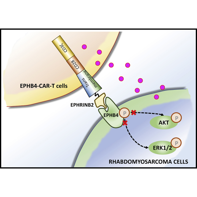
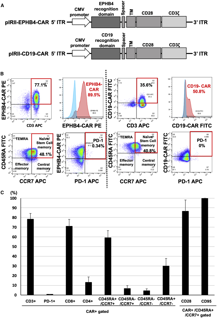
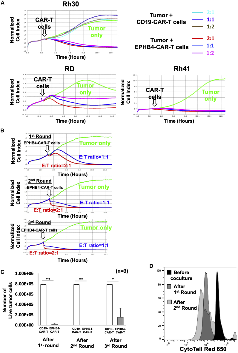
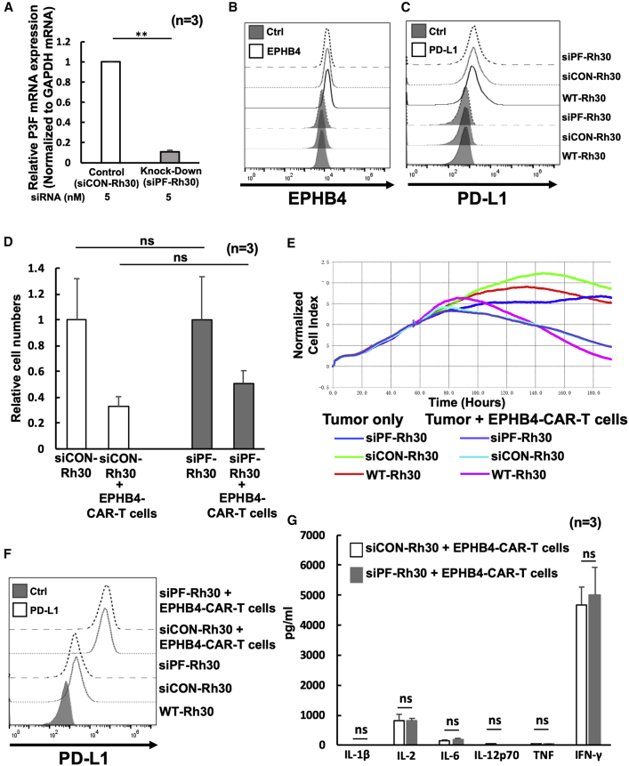
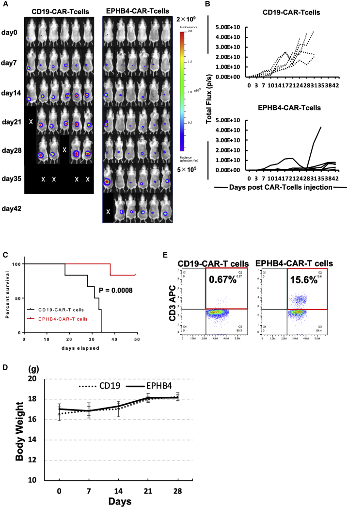
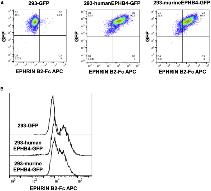
Ephrin type-B receptor 4 (EPHB4), expressed in tumors including rhabdomyosarcoma, is a suitable target for chimeric antigen receptor (CAR)-T cells. Ligand-independent activation of EPHB4 causes cell proliferation and malignant transformation in rhabdomyosarcoma, whereas ligand-dependent stimulation of EPHB4 induces apoptosis in rhabdomyosarcoma. Therefore, we hypothesized that ligand-based, EPHB4-specific CAR-T cells may kill rhabdomyosarcoma cells without stimulating downstream cell proliferation mechanisms. We developed novel CAR-T cells by targeting EPHB4 via EPHRIN B2, a natural ligand of EPHB4. The generation of EPHB4-CAR-T cells via piggyBac (PB) transposon-based gene transfer resulted in sufficient T cell expansion and CAR positivity (78.5% ± 5.9%). PB-EPHB4-CAR-T cells displayed a dominant stem cell memory fraction (59.4% ± 7.2%) as well as low PD-1 expression (0.60% ± 0.21%) after 14 days of expansion. The PB-EPHB4-CAR-T cells inhibited EPHB4-positive tumor cells without activating cell proliferation downstream of EPHB4, even after multiple tumor re-challenges and suppressed tumor growth in xenograft-bearing mice. Therefore, PB-EPHB4-CAR-T cells possess a memory-rich fraction without early T cell exhaustion and show potential as promising therapeutic agents for treating rhabdomyosarcoma and other EPHB4-positive tumors.

Keywords: CAR-T cell therapy; EPHB4; chimeric antigen receptor; piggyBac transposon; rhabdomyosarcoma; stem cell memory-like T cells.


Conflict of interest statement

The authors declare no competing interests.

Figures

None
Graphical abstract
Figure 1
Figure 1
Production and expansion of EPHB4-CAR-T cells by piggyBac-mediated gene transfer and phenotypic analysis (A) Schematic of the EPHB4-CAR construct and CD19-CAR construct. (B) EPHB4-CAR expression, phenotypes, and expression of exhaustion markers on CAR-T cells were assessed via flow cytometry (left). CD19-CAR expression, phenotypes, and expression of exhaustion markers on CAR-T cells were assessed via flow cytometry (right). (C) The phenotype and exhaustion marker of EPHB4-CAR-T cells are represented by the mean ± SD of 5 different donors.
Figure 2
Figure 2
EPHB4-CAR-T cells demonstrated rapid and sustained antitumor activity against EPHB4-positive tumors EPHB4-CAR-T cells, generated by our original method, were co-cultured with EPHB4-positive tumor cells at effector:target (E:T) ratios, and normalized cell index was monitored using a xCELLigence real-time cell analysis. (A) Rh30, Rh41, and RD were co-cultured with EPHB4-CAR-T cells at E:T ratios of 2:1, 1:1, and 1:2 (B and C) Serial tumor rechallenge assay; tumor cell growth was monitored via a xCELLigence real-time cell analysis (B), and the number of live tumor cells 72 h after co-culture with CD19-CAR-T cells or EPHB4-CAR-T cells in each round was measured via flow cytometry (C). Mean ± SD from 3 different experiments are shown. ∗∗p < 0.01. ∗p < 0.05. (D) Cell division of EPHB4-CAR-T cells upon repeated Rh30 cells stimulation.
Figure 3
Figure 3
PAX3-FOXO1 (P3F) fusion gene did not affect the antitumor efficacy of EPHB4-CAR-T cells (A) After transfection with siRNAs against P3F (siPF) into Rh30 cells for 24 h, the knocked-down efficacy of siPF against P3F was assessed by quantitative reverse-transcription polymerase chain reaction (qRT-PCR). ∗∗p < 0.01. (B and C) P3F did not affect EPHB4 (B), and PD-L1 was assessed by flow cytometry (C). (D) Comparison of the antitumor effect in siCON-Rh30 and siPF-Rh30 assessed by flow cytometry. The antitumor effect of the EPHB4-CAR-T cells on the Rh30 cells was evaluated according to the ratio of survival of the Rh30 cells alone to the survival of the Rh30 cells co-cultured with EPHB4-CAR-T cells. Mean ± SD from 3 different experiments are shown. N = not significant. (E) Cell growth of siCON- and siPF-Rh30 co-cultured with EPHB4-CAR-T cells was acquired by a xCELLigence real-time cell analysis. (F) PD-L1 expression between siCON-Rh30 and siPF-Rh30 treated with EPHB4-CAR-T cells was evaluated via flow cytometry. (G) The level of cytokines in the co-culture supernatant siCON-Rh30 or siPF-Rh30 with EPHB4-CAR-T cells for 24 h.
Figure 4
Figure 4
EPHB4-CAR-T cells debulked rhabdomyosarcoma tumors in a murine xenograft model We injected 2 × 106 Rh30-Luc cells into the dorsal area of SCID beige mice. 7 days later, 10 × 106 EPHB4-CAR-T cells or CD19-CAR-T cells (the expression of each CAR molecule was around 80%) were injected into the tail vein of each mouse. (A) Representative bioluminescent images of tumor growth over time in individual mice. (B) Kinetics of Rh30 xenograft progression in individual mice that had received either the CD19 (control) or EPHB4 CAR-T cells by IVIS imaging. (C) Survival analysis of EPHB4-CAR-T cell-treated mice and CD19-CAR-T cell-treated mice (CD19: N = 6; EPHB4: N = 6). (D) Mice treated with EPHB4-CAR-T cells did not show weight loss. (E) Human T cells in peripheral blood of mice on day 31. Representative dot plot data were shown.
Figure 5
Figure 5
Human EPHRIN B2 protein could interact with murine-EPHB4-expressing cells (A and B) 293-GFP, 293-humanEPHB4-GFP, and 293murineEPHB4-GFP were incubated with human EPHRIN B2-Fc chimera protein, and cell-bound EPHRIN B2-Fc was detected by flow cytometry.

References

    1. Porter D.L., Levine B.L., Kalos M., Bagg A., June C.H. Chimeric antigen receptor-modified T cells in chronic lymphoid leukemia. N. Engl. J. Med. 2011;365:725–733. - PMC - PubMed
    1. Brentjens R.J., Davila M.L., Riviere I., Park J., Wang X., Cowell L.G., Bartido S., Stefanski J., Taylor C., Olszewska M. CD19-targeted T cells rapidly induce molecular remissions in adults with chemotherapy-refractory acute lymphoblastic leukemia. Sci. Transl. Med. 2013;5:177ra38. - PMC - PubMed
    1. Brentjens R.J., Rivière I., Park J.H., Davila M.L., Wang X., Stefanski J., Taylor C., Yeh R., Bartido S., Borquez-Ojeda O. Safety and persistence of adoptively transferred autologous CD19-targeted T cells in patients with relapsed or chemotherapy refractory B-cell leukemias. Blood. 2011;118:4817–4828. - PMC - PubMed
    1. Kochenderfer J.N., Dudley M.E., Feldman S.A., Wilson W.H., Spaner D.E., Maric I., Stetler-Stevenson M., Phan G.Q., Hughes M.S., Sherry R.M. B-cell depletion and remissions of malignancy along with cytokine-associated toxicity in a clinical trial of anti-CD19 chimeric-antigen-receptor-transduced T cells. Blood. 2012;119:2709–2720. - PMC - PubMed
    1. Ahmed N., Brawley V., Hegde M., Bielamowicz K., Kalra M., Landi D., Robertson C., Gray T.L., Diouf O., Wakefield A. HER2-Specific Chimeric Antigen Receptor-Modified Virus-Specific T Cells for Progressive Glioblastoma: A Phase 1 Dose-Escalation Trial. JAMA Oncol. 2017;3:1094–1101. - PMC - PubMed