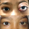
Acute unilateral proptosis in childhood: suspect myeloid sarcoma
- PMID: 33817441
- PMCID: PMC7995499
- DOI: 10.22336/rjo.2021.17
Acute unilateral proptosis in childhood: suspect myeloid sarcoma
Abstract
As the first and only presenting feature of acute myeloid leukemia (AML), unilateral proptosis in children is uncommon. We report the cases of two girls who had no systemic clinical manifestations of AML. Orbital imaging showed space-occupying infiltrating lesions without surrounding bone erosion. Incisional biopsy and immunohistochemistry were diagnostic for myeloid sarcoma. Systemic workup and bone marrow examination showed features of AML. Systemic chemotherapy was administered to both children, who responded well to the treatment. Myeloid sarcoma should be kept in the differentials of the children presenting with isolated proptosis. Immunohistochemistry may provide an accurate diagnosis and early treatment may lead to a prompt recovery with a good prognosis.
Keywords: acute myeloid leukemia; acute proptosis; childhood proptosis; myeloid sarcoma; proptosis.
©Romanian Society of Ophthalmology.
Figures


References
-
- AlSemari MA, Perrotta M, Russo C, Alkatan HM, Maktabi A, Elkhamary S, Crescenzo RMD, Mascolo M, Elefante A, Rombetto L, Capasso R, Strianese D. Orbital myeloid sarcoma (chloroma): Report of 2 cases and literature review. Am J Ophthalmol Case Rep. 2020 Jul 11;19:100806. doi:10.1016/j.ajoc.2020.100806. - PMC - PubMed
-
- Aggarwal E, Mulay K, Honavar SG. Orbital extramedullary granulocytic sarcoma: clinicopathologic correlation with immunohistochemical features. SurvOphthalmol. 2014;59:232–235. - PubMed
-
- Zimmerman LE, Font RL. Ophthalmologic manifestations of granulocytic sarcoma (myeloid sarcoma or chloroma). The third Pan American Association of Ophthalmology and American Journal of Ophthalmology Lecture. Am J Ophthalmol. 1975;80:975–990. - PubMed
-
- Karesh JW, Goldman EJ, Reck K, Kelman SE, Lee EJ, Schiffer CA. A prospective ophthalmic evaluation of patients with acute myeloid leukemia: correlation of ocular and hematologic findings. J Clin Oncol. 1989;7:1528–1532. - PubMed
Publication types
MeSH terms
LinkOut - more resources
Full Text Sources
Other Literature Sources
Research Materials
