Review
. 2021 Mar 6;24(4):102270.
doi: 10.1016/j.isci.2021.102270. eCollection 2021 Apr 23.

Pharmacological and nutritional targeting of voltage-gated sodium channels in the treatment of cancers

Affiliations
Review

Pharmacological and nutritional targeting of voltage-gated sodium channels in the treatment of cancers

Osbaldo Lopez-Charcas et al. iScience. .

Abstract

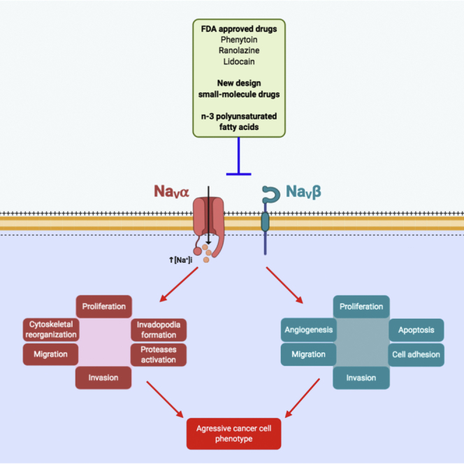
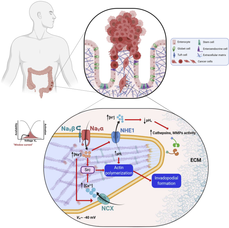
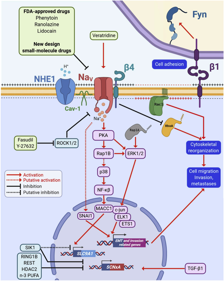
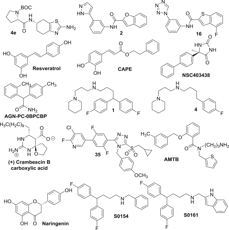
Voltage-gated sodium (NaV) channels, initially characterized in excitable cells, have been shown to be aberrantly expressed in non-excitable cancer tissues and cells from epithelial origins such as in breast, lung, prostate, colon, and cervix, whereas they are not expressed in cognate non-cancer tissues. Their activity was demonstrated to promote aggressive and invasive potencies of cancer cells, both in vitro and in vivo, whereas their deregulated expression in cancer tissues has been associated with metastatic progression and cancer-related death. This review proposes NaV channels as pharmacological targets for anticancer treatments providing opportunities for repurposing existing NaV-inhibitors or developing new pharmacological and nutritional interventions.

Keywords: Cancer; Cell Biology.


Conflict of interest statement

Authors declare no competing interest.

Figures

None
Graphical abstract
Figure 1
Figure 1
Expression of NaVα in carcinoma and role in invadopodial activity and invasion of extracellular matrices Progression of precancerous into cancer cells is illustrated in the context in the malignant transformation of colon epithelium. Transformed cells have lost cell polarity, replication control, and cell-cell adherent junctions, and they acquired a mesenchymal pro-invasive phenotype. Migrating cancer cells develop a specialized actin-based membrane protrusions called “invadopodia” that facilitate cell invasion by providing a coupling of focal extracellular matrix (ECM) degradation together with a directional cell movement. NaV channels are expressed in invadopodial structures, co-localizing with the Na+/H+ exchanger type 1 (NHE1). Activity of NaV channels enhances the extrusion of protons by NHE1 and therefore the acidification of the peri-invadopodial microenvironment, thus favoring both secretion and activity of ECM proteases such as cysteine cathepsins and matrix metalloproteinases (MMPs). Cancer cell resting potential (Vm) is around −40 mV, in a window of voltage of NaV channels (overlap between activation and steady-state inactivation curves) in which a small proportion of channels are activated but non-inactivated, thus generating a small but continuous Na+ influx through a so-called “window sodium current.” NaV channels are also proposed to increase the intracellular levels of Ca2+ ions by the functioning of Na+/Ca2+ exchanger (NCX) in a “reverse mode.” Thus, the increase in the intracellular concentration of Na+ and Ca2+, sustains SRC kinase activity, leading to the polymerization of acting filaments and the formation of invadopodial structure.
Figure 2
Figure 2
Participation of NaVα and/or NaVβ in pro-metastatic signaling pathways NaVα subunit overexpression and activity in cancer cells trigger biochemical or an electro-biochemical cascades, leading to the acquisition of a pro-invasive cell phenotype. NaV is co-localized with NHE1 in caveolin-1 (Cav-1)-containing lipid rafts and promotes the efflux of protons. NaV activity can be further stimulated by the use of pharmacological activators such as veratridine (inhibitor of the inactivation phase). Activity of NaVα subunits leads to a cAMP-independent activation of protein kinase A (PKA) that activates the cytosolic small GTPase Ras-related protein 1 (Rap1A/B) and the extracellular-signal-regulated kinases (ERK1/2). The transcription factor (TF) metastasis associated in colon cancer 1 (MACC1) is activated by the p38/NF-κβ signaling, whereas the TFs c-jun, ELK1, and ETS1 are activated by ERK1/2 and the zinc finger protein SNAI1 is activated through a NaVα-dependent mechanism regulating the expression of genes associated with cytoskeleton reorganization, cell motility, extracellular matrix degradation, and cell invasiveness. It has been demonstrated that MACC1 upregulates the expression of the SLC9A1 gene, encoding for NHE1, thus enhancing its activity at plasma membrane. On the other hand, the electro-biochemical triggering begins with a resting potential depolarization due to the activity of NaVα subunits promoting the activation and recruitment of the small GTPase Ras-related C3 botulinum toxin substrate 1 (Rac1) at the leading edge of migrating cells. Transforming growth factor β 1 (TGF-β1) increases the expression levels of NaV channels genes (SCNxA), whereas ring finger protein 1 (RING1B), RE1 silencing transcription factor (REST), histone deacetylase 2 (HDAC2) and salt inducible kinase 1 (SIK-1), as well as the n-3 polyunsaturated fatty acids n-3 (PUFA) repress their expression. SIK-1 also impairs the functioning of NHE1 exchanger. The “auxiliary subunit” NaVβ4 is expressed in normal epithelial cells but is importantly downregulated in invasive cells and high-grade metastatic tumors. The absence of this protein, but specifically the lack of the intracellular C-terminus domain, triggers the acquisition of an amoeboid-mesenchymal hybrid phenotype dependent of the small GTPase Ras homolog family member A (RhoA). NaVβ1 proteins have a dual role in cancer cells acting as cell adhesion molecules (CAMs), reducing cell migration and proliferation. However, it has also been demonstrated that NaVβ1 promotes tumor growth, metastasis, and vascularization via the proto-oncogene tyrosine-protein kinase Fyn. The Rho-associated protein kinases (ROCK1/2) negatively regulate the expression of NaVα subunits, therefore, silencing or inhibition of these repressors restore NaV channels activity promoting an aggressive cell phenotype. Pharmacological intervention with FDA-approved drugs or new-design small-molecule lead compounds against NaV channels represents a promising strategy to decrease sodium-channel-associated metastases.
Figure 3
Figure 3
Chemical structures of known NaVα blockers with anticancer effects

References

    1. Adachi K., Toyota M., Sasaki Y., Yamashita T., Ishida S., Ohe-Toyota M., Maruyama R., Hinoda Y., Saito T., Imai K. Identification of SCN3B as a novel p53-inducible proapoptotic gene. Oncogene. 2004;23:7791–7798. - PubMed
    1. Agnew W.S., Moore A.C., Levinson S.R., Raftery M.A. Identification of a large molecular weight peptide associated with a tetrodotoxin binding protein from the electroplax of Electrophorus electricus. Biochem. Biophys. Res. Commun. 1980;92:860–866. - PubMed
    1. Agwa A.J., Peigneur S., Chow C.Y., Lawrence N., Craik D.J., Tytgat J., King G.F., Henriques S.T., Schroeder C.I. Gating modifier toxins isolated from spider venom: modulation of voltage-gated sodium channels and the role of lipid membranes. J. Biol. Chem. 2018;293:9041–9052. - PMC - PubMed
    1. Amara S., Ivy M.T., Myles E.L., Tiriveedhi V. Sodium channel gammaENaC mediates IL-17 synergized high salt induced inflammatory stress in breast cancer cells. Cell. Immunol. 2016;302:1–10. - PMC - PubMed
    1. Arnold M., Sierra M.S., Laversanne M., Soerjomataram I., Jemal A., Bray F. Global patterns and trends in colorectal cancer incidence and mortality. Gut. 2017;66:683–691. - PubMed

LinkOut - more resources