Observational Study
. 2021 Mar 19;3(2):e200014.
doi: 10.1148/rycan.2021200014. eCollection 2021 Mar.

The Role of CT in Assessment of Extraregional Lymph Node Involvement in Pancreatic and Periampullary Cancer: A Diagnostic Accuracy Study

Affiliations
Observational Study

The Role of CT in Assessment of Extraregional Lymph Node Involvement in Pancreatic and Periampullary Cancer: A Diagnostic Accuracy Study

Dorine S J Tseng et al. Radiol Imaging Cancer. .

Abstract

Purpose: To investigate the diagnostic accuracy of CT in assessing extraregional lymph node metastases in pancreatic head and periampullary cancer.

Materials and methods: This prospective observational cohort study was performed at two tertiary hepatopancreatobiliary (HPB) referral centers between March 2013 and December 2014. Patients undergoing pancreatoduodenectomy or bypass surgery with or without palliative radiofrequency ablation were included. Extraregional lymph node involvement was defined as positive lymph nodes in the aortocaval window. Two expert HPB radiologists assessed aortocaval lymph nodes at preoperative CT according to a standardized protocol. All tissue from the aortocaval window was collected intraoperatively. Positive histopathologic finding was the reference standard. Analysis of predictive values and diagnostic accuracy was performed.

Results: A total of 198 consecutive patients (mean age, 66 years; range, 39-86 years; 105 men) with pancreatic head or periampullary carcinoma were included. In 70% of patients, a pancreatoduodenectomy was performed, 4% underwent total pancreatectomy, 4% underwent radiofrequency ablation, and 22% underwent bypass surgery. Forty-four patients (22%) had histologically positive aortocaval lymph nodes. Negative predictive value of CT in assessing aortocaval lymph nodes was 80% for both observers, and positive predictive value was 31%-33%. Overall diagnostic accuracy was 69%-70%.

Conclusion: CT has a low diagnostic accuracy in assessing extraregional lymph node metastases in patients suspected of having pancreatic or periampullary cancer.Keywords: CT, Abdomen/GI, Pancreas, Oncology© RSNA, 2021.

Keywords: Abdomen/GI; CT; Oncology; Pancreas.


Conflict of interest statement

Disclosures of Conflicts of Interest: D.S.J.T. disclosed no relevant relationships. B.K.P. disclosed no relevant relationships. M.S.v.L. disclosed no relevant relationships. J.P.P. disclosed no relevant relationships. L.A.B. disclosed no relevant relationships. N.H.M. Activities related to the present article: disclosed no relevant relationships. Activities not related to the present article: institution consults for BMS, MSD, Eli Lilly, Servier, and AstraZeneca; institution is paid by BMS for lectures. Activities related to the present article: disclosed no relevant relationships. Activities not related to the present article: disclosed no relevant relationships. Other relationships: disclosed no relevant relationships. V.d.M. disclosed no relevant relationships. H.C.v.S. disclosed no relevant relationships. J.I.E. disclosed no relevant relationships. I.Q.M. disclosed no relevant relationships.

Figures

Flowchart patient inclusion. FN = false negative, FP = false positive, TN = true negative, TP = true positive.
Figure 1:
Flowchart patient inclusion. FN = false negative, FP = false positive, TN = true negative, TP = true positive.
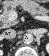
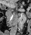
Patient is a 52-year-old woman. Transverse section of 7-mm node (arrow) between aorta (A) and inferior vena cava (IVC), scored as positive based on ill-defined border (true positive).
Figure 2:
Patient is a 52-year-old woman. Transverse section of 7-mm node (arrow) between aorta (A) and inferior vena cava (IVC), scored as positive based on ill-defined border (true positive).
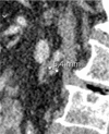
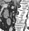
Patient is a 68-year-old woman. Marked differences of minimal node size in the different orthogonal planes: (a) transverse 3.8 mm, (b) sagittal 5.4 mm, and (c) coronal 6.5 mm. Consequently, 4 mm (3.8 rounded to a whole number) was recorded as node size. Node was scored as negative but was a false negative. A = aorta, IVC = inferior vena cava.
Figure 3a:
Patient is a 68-year-old woman. Marked differences of minimal node size in the different orthogonal planes: (a) transverse 3.8 mm, (b) sagittal 5.4 mm, and (c) coronal 6.5 mm. Consequently, 4 mm (3.8 rounded to a whole number) was recorded as node size. Node was scored as negative but was a false negative. A = aorta, IVC = inferior vena cava.
Figure 3b:
Patient is a 68-year-old woman. Marked differences of minimal node size in the different orthogonal planes: (a) transverse 3.8 mm, (b) sagittal 5.4 mm, and (c) coronal 6.5 mm. Consequently, 4 mm (3.8 rounded to a whole number) was recorded as node size. Node was scored as negative but was a false negative. A = aorta, IVC = inferior vena cava.
Figure 3c:
Patient is a 68-year-old woman. Marked differences of minimal node size in the different orthogonal planes: (a) transverse 3.8 mm, (b) sagittal 5.4 mm, and (c) coronal 6.5 mm. Consequently, 4 mm (3.8 rounded to a whole number) was recorded as node size. Node was scored as negative but was a false negative. A = aorta, IVC = inferior vena cava.
Patient is a 79-year-old man. (a) Transverse, (b) coronal, and (c) sagittal images of aortocaval node, with minimal diameters of 3.4 mm, 3.9 mm, and 6.8 mm, respectively. Consequently, 3 mm (3.4 mm rounded to a whole number) was recorded for node size. Node was scored as negative (true negative).
Figure 4a:
Patient is a 79-year-old man. (a) Transverse, (b) coronal, and (c) sagittal images of aortocaval node, with minimal diameters of 3.4 mm, 3.9 mm, and 6.8 mm, respectively. Consequently, 3 mm (3.4 mm rounded to a whole number) was recorded for node size. Node was scored as negative (true negative).
Figure 4b:
Patient is a 79-year-old man. (a) Transverse, (b) coronal, and (c) sagittal images of aortocaval node, with minimal diameters of 3.4 mm, 3.9 mm, and 6.8 mm, respectively. Consequently, 3 mm (3.4 mm rounded to a whole number) was recorded for node size. Node was scored as negative (true negative).
Figure 4c:
Patient is a 79-year-old man. (a) Transverse, (b) coronal, and (c) sagittal images of aortocaval node, with minimal diameters of 3.4 mm, 3.9 mm, and 6.8 mm, respectively. Consequently, 3 mm (3.4 mm rounded to a whole number) was recorded for node size. Node was scored as negative (true negative).
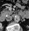
Patient is a 64-year-old man. (a) Transverse and (b) coronal images of node between aorta (A) and inferior vena cava (IVC), with minimal diameter of 11.5 and 10.8 mm, respectively. Because the coronal image yielded the lowest diameter, 11 mm (10.8 rounded to a whole number) was recorded as node size. The node was scored as positive based on size and round shape but was false positive.
Figure 5a:
Patient is a 64-year-old man. (a) Transverse and (b) coronal images of node between aorta (A) and inferior vena cava (IVC), with minimal diameter of 11.5 and 10.8 mm, respectively. Because the coronal image yielded the lowest diameter, 11 mm (10.8 rounded to a whole number) was recorded as node size. The node was scored as positive based on size and round shape but was false positive.
Figure 5b:
Patient is a 64-year-old man. (a) Transverse and (b) coronal images of node between aorta (A) and inferior vena cava (IVC), with minimal diameter of 11.5 and 10.8 mm, respectively. Because the coronal image yielded the lowest diameter, 11 mm (10.8 rounded to a whole number) was recorded as node size. The node was scored as positive based on size and round shape but was false positive.
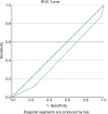
Receiver operating characteristic curve for the model best predicting aortocaval lymph node metastases. Model included tumor growth in surrounding tissue and irregular margins of aortocaval lymph node.
Figure 6:
Receiver operating characteristic curve for the model best predicting aortocaval lymph node metastases. Model included tumor growth in surrounding tissue and irregular margins of aortocaval lymph node.

References

    1. Wagner M, Redaelli C, Lietz M, Seiler CA, Friess H, Büchler MW. Curative resection is the single most important factor determining outcome in patients with pancreatic adenocarcinoma. Br J Surg 2004;91(5):586–594. - PubMed
    1. Tamm EP, Balachandran A, Bhosale PR, et al. . Imaging of pancreatic adenocarcinoma: update on staging/resectability. Radiol Clin North Am 2012;50(3):407–428. - PubMed
    1. Tseng DS, van Santvoort HC, Fegrachi S, et al. . Diagnostic accuracy of CT in assessing extra-regional lymphadenopathy in pancreatic and peri-ampullary cancer: a systematic review and meta-analysis. Surg Oncol 2014;23(4):229–235. - PubMed
    1. Zacharias T, Jaeck D, Oussoultzoglou E, Neuville A, Bachellier P. Impact of lymph node involvement on long-term survival after R0 pancreaticoduodenectomy for ductal adenocarcinoma of the pancreas. J Gastrointest Surg 2007;11(3):350–356. - PubMed
    1. Sohn TA, Yeo CJ, Cameron JL, et al. . Resected adenocarcinoma of the pancreas-616 patients: results, outcomes, and prognostic indicators. J Gastrointest Surg 2000;4(6):567–579. - PubMed

Publication types

LinkOut - more resources