Usefulness of 3-Dimensional-Printed Breast Surgical Guides for Undetectable Ductal Carcinoma In Situ on Ultrasonography: A Report of 2 Cases
- PMID: 33818019
- PMCID: PMC8250104
- DOI: 10.4048/jbc.2021.24.e14
Usefulness of 3-Dimensional-Printed Breast Surgical Guides for Undetectable Ductal Carcinoma In Situ on Ultrasonography: A Report of 2 Cases
Abstract
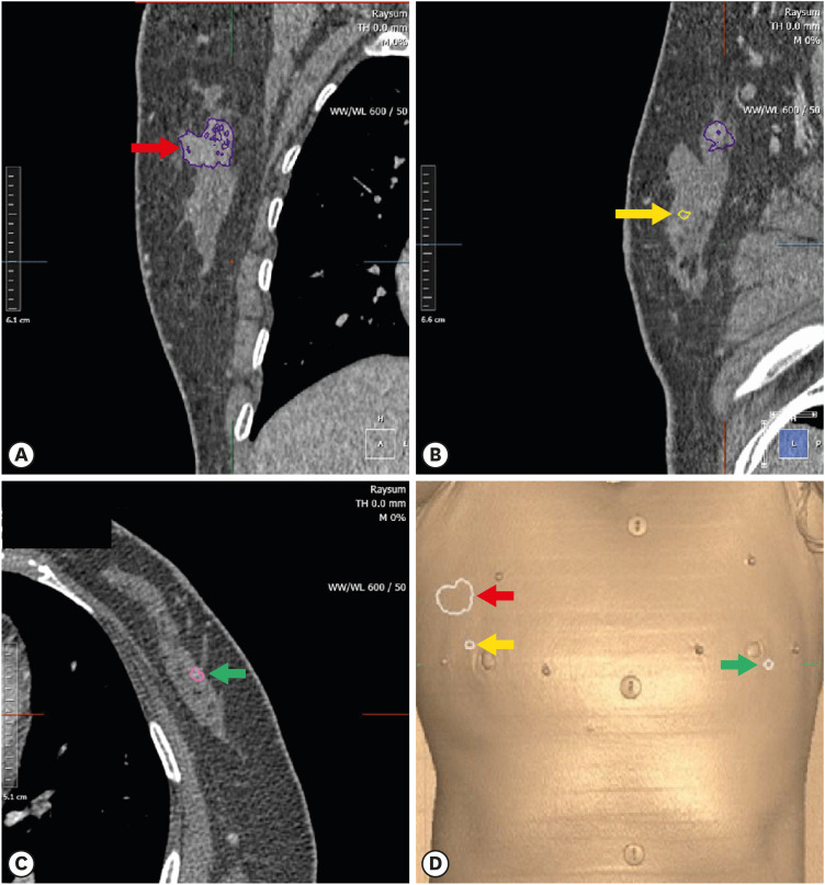
Tumor localization is challenging in the context of ductal carcinoma in situ (DCIS) treated with breast-conserving surgery. Conventional localization methods are generally performed under the guidance of ultrasonography or mammography and are rarely performed with magnetic resonance imaging (MRI), which is more sensitive than the aforementioned modalities in detecting DCIS. Here, we report the application of MRI-based individualized 3-dimensional (3D)-printed breast surgical guides (BSGs) for patients with breast cancer. We successfully resected indeterminate and suspicious lesions that were only detected using preoperative MRI, and the final histopathologic results confirmed DCIS with clear resection margins. MRI guidance combined with 3D-printed BSGs can be used for DCIS localization, especially for lesions easily detectable using MRI only.
Keywords: Breast neoplasms; Carcinoma, ductal; Magnetic resonance imaging; Printing, three-dimensional.
© 2021 Korean Breast Cancer Society.
Conflict of interest statement
BeomSeok Ko and Namkug Kim hold a patent for the 3D-printed breast surgical guide and are the founding members of ANYMEDi, Inc. Zhen-Yu Wu received consulting fees from ANYMEDi, Inc.
Figures


Similar articles
-
Efficacy of breast MRI for surgical decision in patients with breast cancer: ductal carcinoma in situ versus invasive ductal carcinoma.BMC Cancer. 2020 Sep 29;20(1):934. doi: 10.1186/s12885-020-07443-7. BMC Cancer. 2020. PMID: 32993586 Free PMC article.
-
Rates of reexcision for breast cancer after magnetic resonance imaging-guided bracket wire localization.J Am Coll Surg. 2005 Apr;200(4):527-37. doi: 10.1016/j.jamcollsurg.2004.12.013. J Am Coll Surg. 2005. PMID: 15804466
-
Magnetic resonance imaging based 3-dimensional printed breast surgical guide for breast-conserving surgery in ductal carcinoma in situ: a clinical trial.Sci Rep. 2020 Oct 28;10(1):18534. doi: 10.1038/s41598-020-75398-7. Sci Rep. 2020. PMID: 33116237 Free PMC article. Clinical Trial.
-
Magnetic resonance imaging of ductal carcinoma in situ: what is its clinical application? A review.Am J Surg. 2009 Aug;198(2):262-9. doi: 10.1016/j.amjsurg.2009.01.010. Epub 2009 Apr 17. Am J Surg. 2009. PMID: 19375068 Review.
-
Ductal carcinoma in situ of the breast: MR imaging findings with histopathologic correlation.Radiographics. 2010 Oct;30(6):1673-87. doi: 10.1148/rg.306105510. Radiographics. 2010. PMID: 21071382 Review.
Cited by
-
3D and 4D Printing in the Fight against Breast Cancer.Biosensors (Basel). 2022 Jul 26;12(8):568. doi: 10.3390/bios12080568. Biosensors (Basel). 2022. PMID: 35892465 Free PMC article. Review.
-
Radiological Society of North America (RSNA) 3D Printing Special Interest Group (SIG) clinical situations for which 3D printing is considered an appropriate representation or extension of data contained in a medical imaging examination: breast conditions.3D Print Med. 2023 Mar 23;9(1):8. doi: 10.1186/s41205-023-00171-1. 3D Print Med. 2023. PMID: 36952139 Free PMC article.
References
-
- Siegel RL, Miller KD, Jemal A. Cancer statistics, 2019. CA Cancer J Clin. 2019;69:7–34. - PubMed
-
- Berg WA, Gutierrez L, NessAiver MS, Carter WB, Bhargavan M, Lewis RS, et al. Diagnostic accuracy of mammography, clinical examination, US, and MR imaging in preoperative assessment of breast cancer. Radiology. 2004;233:830–849. - PubMed
-
- Kuhl CK, Schrading S, Bieling HB, Wardelmann E, Leutner CC, Koenig R, et al. MRI for diagnosis of pure ductal carcinoma in situ: a prospective observational study. Lancet. 2007;370:485–492. - PubMed
-
- Gray RJ, Salud C, Nguyen K, Dauway E, Friedland J, Berman C, et al. Randomized prospective evaluation of a novel technique for biopsy or lumpectomy of nonpalpable breast lesions: radioactive seed versus wire localization. Ann Surg Oncol. 2001;8:711–715. - PubMed
Publication types
Grants and funding
LinkOut - more resources
Full Text Sources
Other Literature Sources