. 2021 Apr 27;15(4):6562-6572.
doi: 10.1021/acsnano.0c09732. Epub 2021 Apr 5.

Lipophilicity of Cationic Ligands Promotes Irreversible Adsorption of Nanoparticles to Lipid Bilayers

Affiliations

Lipophilicity of Cationic Ligands Promotes Irreversible Adsorption of Nanoparticles to Lipid Bilayers

Christian A Lochbaum et al. ACS Nano. .

Abstract

A mechanistic understanding of the influence of the surface properties of engineered nanomaterials on their interactions with cells is essential for designing materials for applications such as bioimaging and drug delivery as well as for assessing nanomaterial safety. Ligand-coated gold nanoparticles have been widely investigated because their highly tunable surface properties enable investigations into the effect of ligand functionalization on interactions with biological systems. Lipophilic ligands have been linked to adverse biological outcomes through membrane disruption, but the relationship between ligand lipophilicity and membrane interactions is not well understood. Here, we use a library of cationic ligands coated on 2 nm gold nanoparticles to probe the impact of ligand end group lipophilicity on interactions with supported phosphatidylcholine lipid bilayers as a model for cytoplasmic membranes. Nanoparticle adsorption to and desorption from the model membranes were investigated by quartz crystal microbalance with dissipation monitoring. We find that nanoparticle adsorption to model membranes increases with ligand lipophilicity. The effects of ligand structure on gold nanoparticle attachment were further analyzed using atomistic molecular dynamics simulations, which showed that the increase in ligand lipophilicity promotes ligand intercalation into the lipid bilayer. Together, the experimental and simulation results could be described by a two-state model that accounts for the initial attachment and subsequent conversion to a quasi-irreversibly bound state. We find that only nanoparticles coated with the most lipophilic ligands in our nanoparticle library undergo conversion to the quasi-irreversible state. We propose that the initial attachment is governed by interaction between the ligands and phospholipid tail groups, whereas conversion into the quasi-irreversibly bound state reflects ligand intercalation between phospholipid tail groups and eventual lipid extraction from the bilayer. The systematic variation of ligand lipophilicity enabled us to demonstrate that the lipophilicity of cationic ligands correlates with nanoparticle-bilayer adsorption and suggested that changing the nonpolar ligand R group promotes a mechanism of ligand intercalation into the bilayer associated with irreversible adsorption.

Keywords: classical molecular dynamics simulations; ligand-coated gold nanoparticles; nanobio interface; quartz crystal microbalance with dissipation monitoring; structure−property relationship; supported lipid bilayers; umbrella sampling.


Conflict of interest statement

The authors declare no competing interests.

Figures

Figure 1.
Figure 1.
Experimental and computational systems used to study gold nanoparticle adsorption onto phospholipid bilayers. (a) Ligands are comprised of an alkane group (gray), an oligo(ethylene glycol) spacer group (green), and a cationic quaternary ammonium group (red) substituted with the indicated R group and two methyl groups. The five R groups used are displayed in red and labeled with their calculated log Klip-w values in parentheses. (b) Schematic of the system used in quartz crystal microbalance experiments to measure nanoparticle adsorption to supported DOPC lipid bilayers. (c) Snapshot of 2-nm gold nanoparticle with C10 ligands placed above a DOPC lipid bilayer. The color scheme is illustrated for each of the components at right. The DOPC lipids are comprised of a zwitterionic phosphatidylcholine head group and nonpolar acyl tails consisting primarily of aliphatic carbon atoms. Water is shown in grey.
Figure 2.
Figure 2.
Influence of ligand lipophilicity on AuNP attachment to supported DOPC bilayers as determined by QCM-D. (a) Acoustic surface mass density (Γ) maximum and after rinse. (b) Dissipation factor (ΔD) maximum and after rinse. Error bars represent one standard deviation from four replicate QCM-D experiments.
Figure 3.
Figure 3.
Free energy as a function of the z distance between the AuNP and lipid bilayer. Potential mean force (PMF) versus z for C1− and C10-AuNPs when the gold core is (a) pulled towards (i.e. decreasing-z) and (b) away from (i.e. increasing-z) the DOPC lipid bilayer. Simulation snapshots show the last configuration from umbrella sampling simulations of C10-AuNPs for different values of z. Water and chlorine atoms are omitted for clarity. Legends are the same for (a) and (b). (c) Number of hydrophobic contacts (ch) versus z for both decreasing- and increasing-z simulations. Hydrophobic contacts are defined as the number of contacts between nonpolar groups in the ligands and in the DOPC tail groups. Error bars are reported as the standard deviation between two 20 ns blocks for each umbrella sampling window.
Figure 4.
Figure 4.
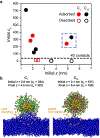
Unbiased simulations initiated from umbrella sampling trajectories. (a) Number of hydrophobic contacts (ch) versus z for unbiased simulations initiated from increasing- and decreasing-z US configurations for C1− and C10-AuNPs. AuNPs are considered adsorbed if z < 6 nm for the last 10 ns of the unbiased simulation (filled markers) and desorbed if z > 6 nm (hollow markers). Points in the dashed blue box were for unbiased simulations initiated from increasing-z US configuration, all other points were for simulations initiated from decreasing-z US configurations. (b) Simulation snapshots after 50 ns of unbiased simulation for C1− and C10-AuNPs. The initial z values and final z values after 50 ns are labeled above the snapshots. Atoms in the DOPC head groups are omitted for clarity.
Figure 5.
Figure 5.
Free energy as a function of AuNP-bilayer hydrophobic contacts. (a) Potential of mean force (PMF) versus the number of hydrophobic contacts (ch) for C1−, Bn-, and C10-AuNPs. Error bars are reported as the standard deviation between two 30 ns trajectories for C1− and C10-AuNPs and two 20 ns trajectories for Bn-AuNP in each umbrella sampling window. (b) Simulation snapshots with ch = 5, 40, and 150 for C1−, Bn-, and C10-AuNPs. DOPC lipids that are within 0.35 nm of the ligand atoms are highlighted in cyan.
Figure 6.
Figure 6.
The role of ligand end group lipophilicity on adsorption to and desorption from phospholipid bilayers. (a) Adsorption rate constant (ka) calculated from AuNP adsorption with calculated desorption rate constant (kd) values. (b) Rate constants for conversion to quasi-irreversibly adsorbed state (kβ) calculated from mass at maximum and after rinse. (c) Desorption rate constants (kd) calculated from AuNP desorption. Error bars represent one standard deviation of four replicate QCM-D measurements. (d) Schematic showing hypothesized mechanism for preferential adsorption of C10-AuNPs compared to C1-AuNPs. C10-AuNPs have a longer R group, denoted by the red lines. Alkane is shown as black lines and PEG is shown as green lines. The symbol and color for the gold core and lipid bilayer is the same as Figure 1b.

References

    1. Maurer-Jones MA; Gunsolus IL; Murphy CJ; Haynes CL Toxicity of Engineered Nanoparticles in the Environment. Anal. Chem 2013, 85 (6), 3036–3049. 10.1021/ac303636s. - DOI - PMC - PubMed
    1. Nel A; Xia T; Mädler L; Li N Toxic Potential of Materials at the Nanolevel. Science. 2006, 311 (5761), 622–627. 10.1126/science.1114397. - DOI - PubMed
    1. Hamers RJ Nanomaterials and Global Sustainability. Acc. Chem. Res 2017, 50 (3), 633–637. 10.1021/acs.accounts.6b00634. - DOI - PubMed
    1. Johnston LJ; Gonzalez-Rojano N; Wilkinson KJ; Xing B Key Challenges for Evaluation of the Safety of Engineered Nanomaterials. NanoImpact 2020, 18, 100219. 10.1016/j.impact.2020.100219. - DOI
    1. Zhu W; von dem Bussche A; Yi X; Qiu Y; Wang Z; Weston P; Hurt RH; Kane AB; Gao H Nanomechanical Mechanism for Lipid Bilayer Damage Induced by Carbon Nanotubes Confined in Intracellular Vesicles. Proc. Natl. Acad. Sci 2016, 113 (44), 12374–12379. 10.1073/pnas.1605030113. - DOI - PMC - PubMed

Publication types

LinkOut - more resources