

Comparison of the histopathological differences between the spinal material and posterior longitudinal ligament in patients with lumbar disc herniation: A focus on the etiopathogenesis
- PMID: 33818148
- PMCID: PMC8020649
- DOI: 10.5144/0256-4947.2021.115
Comparison of the histopathological differences between the spinal material and posterior longitudinal ligament in patients with lumbar disc herniation: A focus on the etiopathogenesis
Abstract
Background: Lumbar disc herniation (LDH) occurs owing to the inability of the posterior longitudinal ligament (PLL) to preserve the disc material within the intervertebral space. There is apparently no study that has investigated the histopathological changes occurring in both PLL and disc material in patients with LDH.
Objective: Investigate and compare the histopathological changes occurring in PLL and disc material of the patients who underwent a surgical operation for LDH.
Design: Descriptive, cross-sectional.
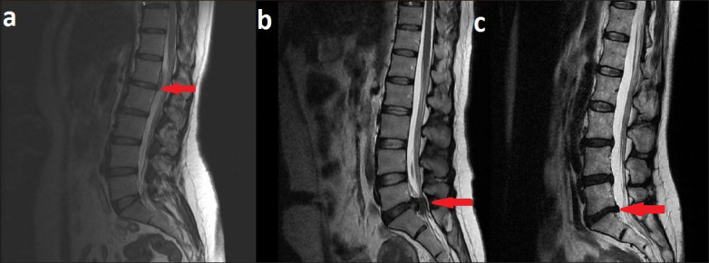
Setting: Pathology and neurosurgery departments of a tertiary health care institution PATIENTS AND METHODS: The study included patients who underwent surgical operation for LDH from January 2018 to May 2019 and whose PLL and disc material were removed together, and had disc degeneration findings that were radiologically and histologically concordant.
Main outcome measures: PLL degeneration scores according to the histopathological findings, changes in disc materials according to the MRI findings, disc degeneration scores according to the histo-pathological findings.
Sample size: 50.
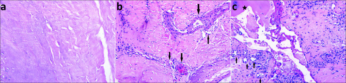
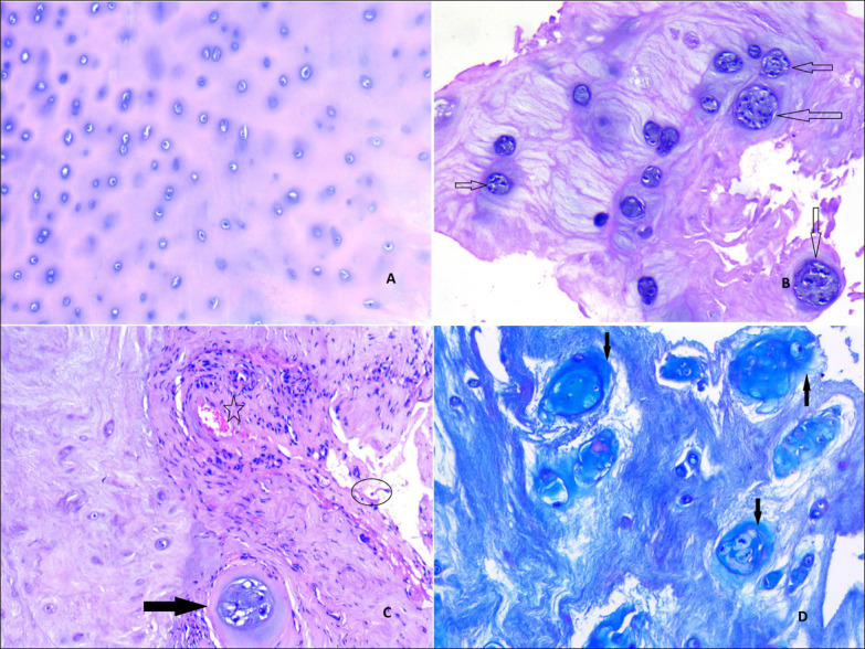
Results: MRI and histological examinations showed fully degenerated black discs (Grade 2) in 12 patients, partially degenerated discs (Grade 1) in 29 patients and fresh/acute discs (Grade 0) in 9 patients. The PLL showed grade 0 degeneration in 2 patients, grade 1 degeneration in 23 patients, and grade 2 degeneration in 25 patients. PLL degeneration grades were higher than the disc degeneration grades (P=.002).
Conclusion: Longitudinal ligament degeneration can play a significant role in the pathogenesis of LDH. To the best of our knowledge, this study represents the first to focus on the histopathological changes occurring in both the PLL and disc material in patients with LDH.
Limitations: Small sample, retrospective CONFLICT OF INTEREST: None.
Figures
Similar articles
-
Differences in calcification and osteogenic potential of herniated discs according to the severity of degeneration based on Pfirrmann grade: a cross-sectional study.BMC Musculoskelet Disord. 2016 Apr 29;17:191. doi: 10.1186/s12891-016-1015-x. BMC Musculoskelet Disord. 2016. PMID: 27495942 Free PMC article.
-
Imaging characteristics of intradural disc herniation: A comparison with large disc extrusion.Eur J Radiol. 2021 Apr;137:109569. doi: 10.1016/j.ejrad.2021.109569. Epub 2021 Jan 27. Eur J Radiol. 2021. PMID: 33578086
-
Lumbar Disc Herniation is a Nonnegligible Factor for the Degeneration of Sacroiliac Joints.Pain Physician. 2021 May;24(3):E357-E365. Pain Physician. 2021. PMID: 33988958
-
Relationships between paraspinal muscle morphology and neurocompressive conditions of the lumbar spine: a systematic review with meta-analysis.BMC Musculoskelet Disord. 2018 Sep 27;19(1):351. doi: 10.1186/s12891-018-2266-5. BMC Musculoskelet Disord. 2018. PMID: 30261870 Free PMC article.
-
Mechanisms and management of self-resolving lumbar disc herniation: bridging molecular pathways to non-surgical clinical success.J Orthop Surg Res. 2025 May 27;20(1):528. doi: 10.1186/s13018-025-05959-x. J Orthop Surg Res. 2025. PMID: 40426259 Free PMC article. Review.
Cited by
-
Posterior longitudinal ligament suturation after lumbar discectomy provides postoperative a large intradural area: First report.J Craniovertebr Junction Spine. 2023 Apr-Jun;14(2):181-186. doi: 10.4103/jcvjs.jcvjs_10_23. Epub 2023 Jun 13. J Craniovertebr Junction Spine. 2023. PMID: 37448510 Free PMC article.
-
Role of Posterior Longitudinal Ligament Complex in Spinal Deformity Secondary to Surgical Resection of the Intradural Tumor.Orthop Surg. 2023 Mar;15(3):819-828. doi: 10.1111/os.13636. Epub 2023 Jan 31. Orthop Surg. 2023. PMID: 36720712 Free PMC article.
References
-
- Wu JC, Mummaneni PV. Lumbar disc herniation and surgical management. World neurosurg. 2010;74(6):572-3. - PubMed
-
- Greenberg MS. Handbook of Neurosurgery. 6th edition. Thieme medical publisher; 2006;pp:289-312.
-
- Pytel P, Wollmann RL, Fessler RG, Krausz TN, Montag AG. Degenerative spine disease: pathologic findings in 985 surgical specimens. Am J Clin Pathol. 2006;125(2):193-202. - PubMed
MeSH terms
LinkOut - more resources
Full Text Sources
Other Literature Sources
Medical
Research Materials