. 2021 Nov;16(11):2243-2249.
doi: 10.4103/1673-5374.310689.

Teriflunomide provides protective properties after oxygen-glucose-deprivation in hippocampal and cerebellar slice cultures

Affiliations

Teriflunomide provides protective properties after oxygen-glucose-deprivation in hippocampal and cerebellar slice cultures

Anna Wolters et al. Neural Regen Res. 2021 Nov.

Abstract

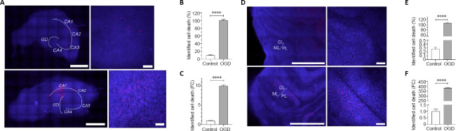
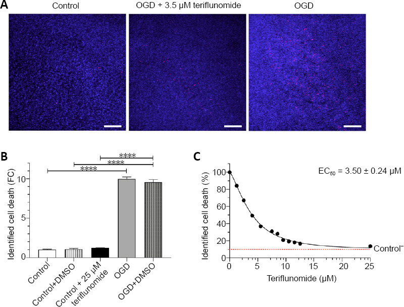
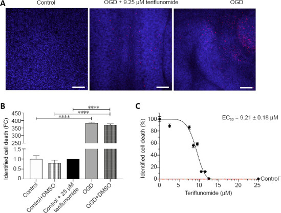
One of the major challenges in emergency medicine is out-of-hospital cardiac arrest (OHCA). Every year, about 53-62/100 000 people worldwide suffer an out-of-hospital cardiac arrest with serious consequences, whereas persistent brain injury is a major cause of morbidity and mortality of those surviving a cardiac arrest. Today, only few and insufficient strategies are known to limit neurological damage of ischemia and reperfusion injury. The aim of the present study was to investigate whether teriflunomide, an approved drug for treatment of relapsing-remitting-multiple-sclerosis, exerts a protective effect on brain cells in an in vitro model of ischemia. Therefore, organotypic slice cultures from rat hippocampus and cerebellum were exposed to oxygen-glucose-deprivation and subsequently treated with teriflunomide. The administration of teriflunomide in the reperfusion time on both hippocampal and cerebellar slice cultures significantly decreased the amount of detectable propidium iodide signal compared with an untreated culture, indicating that more cells survive after oxygen-glucose-deprivation. However, hippocampal slice cultures showed a higher vulnerability to ischemic conditions and a more sensitive response to teriflunomide compared with cerebellar slice cultures. Our study suggests that teriflunomide, applied as a post-treatment after an oxygen-glucose-deprivation, has a protective effect on hippocampal and cerebellar cells in organotypic slice cultures of rats. All procedures were conducted under established standards of the German federal state of North Rhine Westphalia, in accordance with the European Communities Council Directive 2010/63/EU on the protection of animals used for scientific purposes.

Keywords: brain damage; cardiac arrest; cell death; hypoxic chamber; ischemia; organotypic slice cultures; post-treatment; resuscitation.


Conflict of interest statement

None

Figures

Figure 1
Figure 1
A 30-minute OGD induces identifiable cell death in hippocampal and cerebellar organotypic slice cultures. (A) Representative images of hippocampal organotypic slice cultures. Shown are control (upper images) and OGD group (lower images). Cell nuclei were stained with DAPI (blue). Identification of dead cells was performed by staining with propidium iodide (red). Pictures were taken using a confocal spinning disc microscope with a 20× objective. Staining showed increased PI-signal in rat hippocampus after 30-minute OGD. PI-positive cells were found mainly in CA1 and the dentate gyrus (gyrus dentatus = GD). Left image: Scale bar: 1000 µm. Right image: Scale bar: 100 µm. (B) Identified cell death in the organotypic slice cultures of the hippocampus in control and OGD group. PI-signal was normalized to DAPI-signal - OGD-treated group was set to 100 % of identified cell death. (C) Identified cell death calculated as fold change [FC] to control conditions. PI-signal was normalized to DAPI-signal. (D) Representative images of cerebellar organotypic slice cultures. Shown are control (upper images) and OGD group (lower images). Cell nuclei were stained with DAPI (blue). Identification of dead cells was performed by staining with propidium iodide (red). Pictures were taken using a confocal spinning disc microscope with a 20× objective. Staining showed increased PI-signal in rat cerebellum after 30-minute OGD. Damage was identified as multiple diffuse cell nests mainly localized in granular layer (GL) and molecular layer (ML). Purkinje cell layer (PL) showed no PI positive signals after OGD. Left image: Scale bar: 1000 µm. Right image: Scale bar: 100 µm. (E) Identified cell death in the organotypic slice cultures of the cerebellum in control and OGD. PI-signal was normalized to DAPI-signal-OGD-treated group was set to 100 % of identified cell death. (F) Identified cell death calculated as fold change [FC] to control conditions. PI-signal was normalized to DAPI-signal. Data are shown as mean ± SEM; ****P < 0.0001 (two-tailed Student’s t-test); (B, C) n = 13–26 slice cultures per group, three independent preparations; (E, F) n = 9–18 slice cultures per group, three independent preparations. DAPI: 4′,6-Diamidin-2-phenylindol; OGD: oxygen-glucose-deprivation.
Figure 2
Figure 2
Protective effect of teriflunomide on cells of hippocampal organotypic slice cultures after 30-minutes of OGD. (A) Representative image of the CA1 area of a hippocampal organotypic slice culture. Shown are the control group (left), an organotypic slice culture after 30 minutes of OGD (middle), and an OGD group after treatment with the half maximal effective concentration (EC50) of teriflunomide [EC50 = 3.53 ± 0.14 µM]. The cell nuclei were stained with DAPI (blue). The identification of dead cells with a permeable membrane was performed by staining with propidium iodide (red). Pictures were taken using a confocal spinning disc microscope with a 20× objective. Treatment of the OGD group with teriflunomide leads to a significant reduction of the identified cell death in CA1 and dentate gyrus. Scale bars: 100 µm. (B) Identified cell death in the hippocampal slice cultures after treatment with different concentrations of teriflunomide. PI-signal was normalized to DAPI-signal. Identified cell death was calculated as fold change (FC) to control conditions. The application of DMSO or 25 µM teriflunomide had no effect on the identified cell death under control conditions. After performing OGD, DMSO has no effect on the identified cell death. (C) Concentration dependence of teriflunomide on identified cell death after 30-minute OGD. PI-signal was normalized to DAPI-signal and OGD-treated group was set to 100 % of identified cell death. The identified cell death [%] is significantly reduced with increasing drug concentration of the administered teriflunomide. Data are shown as mean ± SEM; ****P < 0.0001 (one-way analysis of variance with Tukey’s multiple comparisons post hoc test); n = 3 (B) and 9–26 (C) slice cultures per group, three independent preparations. OGD: Oxygen-glucose-deprivation.
Figure 3
Figure 3
Protective effect of teriflunomide on cells of cerebellar organotypic slice cultures after 30-minutes of OGD. (A) Representative image of in granular layer and molecular layer of a cerebellar organotypic slice culture. Shown are the control group (left), an organotypic slice culture after 30 minutes of OGD (middle), and an OGD group after treatment with the half maximal effective concentration (EC50) of teriflunomide [EC50 = 9.22 ± 0.15 µM]. The cell nuclei were stained with DAPI (blue). The identification of dead cells with a permeable membrane was performed by staining with propidium iodide (red). Pictures were taken using a confocal spinning disc microscope with a 20× objective. The staining showed increased cell death in the rat cerebellar slice cultures after 30-minute of OGD. PI-positive cells were identified as multiple diffuse cell nests mainly in granular and molecular layer. Treatment of the OGD group with teriflunomide leads to a significant reduction of the identified cell death. Scale bars: 100 µm. (B) Identified cell death in the cerebellar slice cultures after treatment with different concentrations of teriflunomide. PI-signal was normalized to DAPI-signal. Identified cell death was calculated as fold change (FC) to control conditions. The application of DMSO or 25 µM teriflunomide had no effect on the identified cell death under control conditions. After performing OGD, DMSO has no effect on the identified cell death. (C) Concentration dependence of teriflunomide on identified cell death after 30-minute OGD. PI-signal was normalized to DAPI-signal and OGD-treated group was set to 100 % of identified cell death. The identified cell death is significantly reduced with increasing drug concentration of the administered teriflunomide. Data are shown as mean ± SEM; ****P < 0.0001 (one-way analysis of variance with Tukey’s multiple comparisons post hoc test); n = 3 (B) and 10–21 (C) slice cultures per group, three independent preparations. OGD: Oxygen-glucose-deprivation; PI: propidium iodide.

Similar articles

Cited by

References

    1. Ambrosius B, Faissner S, Guse K, von Lehe M, Grunwald T, Gold R, Grewe B, Chan A. Teriflunomide and monomethylfumarate target HIV-induced neuroinflammation and neurotoxicity. J Neuroinflammation. 2017;14:51. - PMC - PubMed
    1. Arawwawala D, Brett SJ. Clinical review: Beyond immediate survival from resuscitation - Long-term outcome considerations after cardiac arrest. Crit Care. 2007;11:235. - PMC - PubMed
    1. Bar-Or A. Teriflunomide (Aubagio®) for the treatment of multiple sclerosis. Exp Neurol. 2014;262:57–65. - PubMed
    1. Berdowski J, Berg RA, Tijssen JGP, Koster RW. Global incidences of out-of-hospital cardiac arrest and survival rates: Systematic review of 67 prospective studies. Resuscitation. 2010;81:1479–1487. - PubMed
    1. Bernard SA, Gray TW, Buist MD, Jones BM, Silvester W, Gutteridge G, Smith K. Treatment of comatose survivors of out-of-hospital cardiac arrest with induced hypothermia. N Engl J Med. 2002;346:557–563. - PubMed