

Apelin-13 regulates electrical activity in the globus pallidus and induces postural changes in rats
- PMID: 33818511
- PMCID: PMC8354122
- DOI: 10.4103/1673-5374.310694
Apelin-13 regulates electrical activity in the globus pallidus and induces postural changes in rats
Abstract
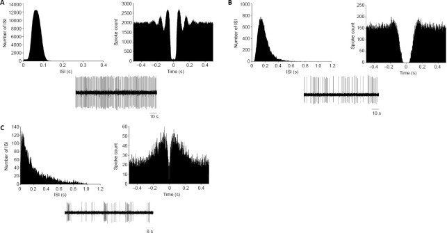
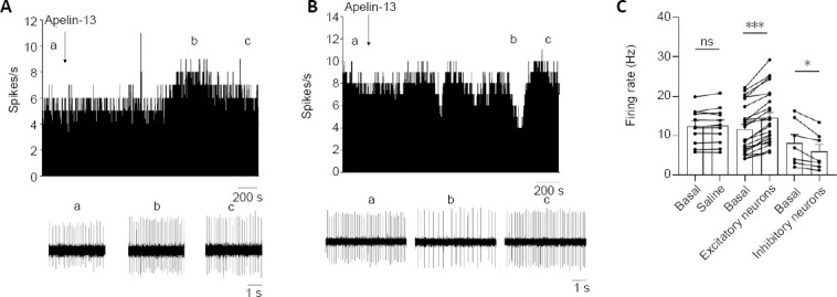
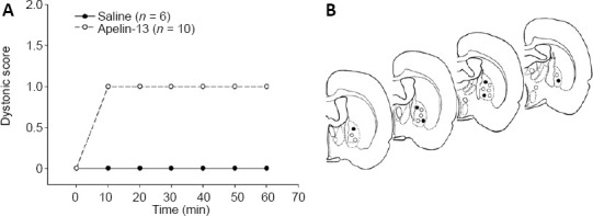
The globus pallidus is the relay nucleus of the basal ganglia, and changes in its electrical activity can cause motor impairment. Apelin-13 is widely distributed in the central and peripheral nervous systems. It has been demonstrated that apelin-13 plays important roles in the regulation of blood pressure and other non-motor functions. However, its role in motor function has rarely been reported. In the present study, apelin-13 (10 μM/100 μM) was injected into the globus pallidus of rats. The results showed that apelin-13 increased the spontaneous discharges in the majority of pallidal neurons. However, an apelin-13-induced inhibitory effect on the firing rate was also observed in a few pallidal neurons. In postural tests, after the systemic administration of haloperidol, unilateral pallidal injection of apelin-13 caused a contralateral deflection. Together, these findings suggest that apelin-13 regulates the electrical activity of pallidal neurons and thus participates in central motor control in rats. The study was approved by the Animal Ethics Committee of Qingdao University (approval No. 20200615Wistar0451003020) on June 15, 2020.
Keywords: apelin-13; basal ganglion; electrophysiology; firing rate; globus pallidus; microinjection; motor behavior; movement disorder.
Conflict of interest statement
None
Figures
Similar articles
-
Orexin-A increases the activity of globus pallidus neurons in both normal and parkinsonian rats.Eur J Neurosci. 2016 Sep;44(5):2247-57. doi: 10.1111/ejn.13323. Epub 2016 Jul 13. Eur J Neurosci. 2016. PMID: 27336845
-
Electrophysiological and behavioral effects of neurotensin in rat globus pallidus: an in vivo study.Exp Neurol. 2007 May;205(1):108-15. doi: 10.1016/j.expneurol.2007.01.031. Epub 2007 Feb 13. Exp Neurol. 2007. PMID: 17362935
-
In vivo Bidirectional Modulation of Cannabinoid on the Activity of Globus Pallidus in Rats.Neuroscience. 2021 Aug 1;468:123-138. doi: 10.1016/j.neuroscience.2021.06.012. Epub 2021 Jun 12. Neuroscience. 2021. PMID: 34129911
-
The functions of the basal ganglia and the paradox of stereotaxic surgery in Parkinson's disease.Brain. 1994 Aug;117 ( Pt 4):877-97. doi: 10.1093/brain/117.4.877. Brain. 1994. PMID: 7922472 Review.
-
The globus pallidus as a target for neuropeptides and endocannabinoids participating in central activities.Peptides. 2020 Feb;124:170210. doi: 10.1016/j.peptides.2019.170210. Epub 2019 Nov 26. Peptides. 2020. PMID: 31778724 Review.
Cited by
-
Beneficial effects of Apelin-13 on metabolic diseases and exercise.Front Endocrinol (Lausanne). 2023 Nov 28;14:1285788. doi: 10.3389/fendo.2023.1285788. eCollection 2023. Front Endocrinol (Lausanne). 2023. PMID: 38089606 Free PMC article. Review.
-
The Mechanism of the Nucleus Accumbens-Ventral Pallidum Pathway Mediated by Drug Withdrawal-Induced High-Seeking Motivation in Cocaine Addiction.Int J Mol Sci. 2024 Oct 29;25(21):11612. doi: 10.3390/ijms252111612. Int J Mol Sci. 2024. PMID: 39519163 Free PMC article.
References
-
- Abecassis ZA, Berceau BL, Win PH, García D, Xenias HS, Cui Q, Pamukcu A, Cherian S, Hernández VM, Chon U, Lim BK, Kim Y, Justice NJ, Awatramani R, Hooks BM, Gerfen CR, Boca SM, Chan CS. Npas1(+)-Nkx2.1(+) neurons are an integral part of the cortico-pallido-cortical loop. J Neurosci. 2020;40:743–768. - PMC - PubMed
-
- Antushevich H, Wójcik M. Review: apelin in disease. Clin Chim Acta. 2018;483:241–248. - PubMed
-
- Choe W, Albright A, Sulcove J, Jaffer S, Hesselgesser J, Lavi E, Crino P, Kolson DL. Functional expression of the seven-transmembrane HIV-1 co-receptor APJ in neural cells. J Neurovirol 6 Suppl. 2000;1:S61–69. - PubMed
-
- Clarke KJ, Whitaker KW, Reyes TM. Diminished metabolic responses to centrally-administered apelin-13 in diet-induced obese rats fed a high-fat diet. J Neuroendocrinol. 2009;21:83–89. - PubMed
LinkOut - more resources
Full Text Sources
Other Literature Sources
