

Transcriptome analysis of molecular mechanisms underlying facial nerve injury repair in rats
- PMID: 33818518
- PMCID: PMC8354104
- DOI: 10.4103/1673-5374.310700
Transcriptome analysis of molecular mechanisms underlying facial nerve injury repair in rats
Abstract
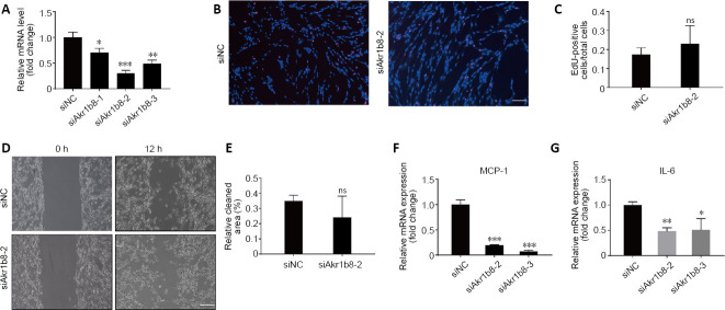
Although the transcriptional alterations inside the facial nucleus after facial nerve injury have been well studied, the gene expression changes in the facial nerve trunk after injury are still unknown. In this study, we established an adult rat model of facial nerve crush injury by compressing the right lateral extracranial nerve trunk. Transcriptome sequencing, differential gene expression analysis, and cluster analysis of the injured facial nerve trunk were performed, and 39 intersecting genes with significant variance in expression were identified. Gene Ontology annotation and Kyoto Encyclopedia of Genes and Genomes pathway analyses of the 39 intersecting genes revealed that these genes are mostly involved in leukocyte cell-cell adhesion and phagocytosis and have essential roles in regulating nerve repair. Quantitative real-time polymerase chain reaction assays were used to validate the expression of pivotal genes. Finally, nine pivotal genes that contribute to facial nerve recovery were identified, including Arhgap30, Akr1b8, C5ar1, Csf2ra, Dock2, Hcls1, Inpp5d, Sla, and Spi1. Primary Schwann cells were isolated from the sciatic nerve of neonatal rats. After knocking down Akr1b8 in Schwann cells with an Akr1b8-specific small interfering RNA plasmid, expression levels of monocyte chemoattractant protein-1 and interleukin-6 were decreased, while cell proliferation and migration were not obviously altered. These findings suggest that Akr1b8 likely regulates the interaction between Schwann cells and macrophages through regulation of cytokine expression to promote facial nerve regeneration. This study is the first to reveal a transcriptome change in the facial nerve trunk after facial nerve injury, thereby revealing the potential mechanism underlying repair of facial nerve injury. This study was approved by the Animal Ethics Committee of Nantong University, China in 2018 (approval No. S20180923-007).
Keywords: Akr1b8; Gene-Act Networks; RNA-Seq; Schwann cells; cell proliferation; facial nerve injury; inflammatory response; transcriptomics analysis.
Conflict of interest statement
None
Figures


Similar articles
-
A Schwann cell-enriched circular RNA circ-Ankib1 regulates Schwann cell proliferation following peripheral nerve injury.FASEB J. 2019 Nov;33(11):12409-12424. doi: 10.1096/fj.201900965R. Epub 2019 Sep 16. FASEB J. 2019. PMID: 31415184 Free PMC article.
-
Identification of key genes involved in axon regeneration and Wallerian degeneration by weighted gene co-expression network analysis.Neural Regen Res. 2022 Apr;17(4):911-919. doi: 10.4103/1673-5374.322473. Neural Regen Res. 2022. PMID: 34472493 Free PMC article.
-
The long noncoding RNA Arrl1 inhibits neurite outgrowth by functioning as a competing endogenous RNA during neuronal regeneration in rats.J Biol Chem. 2020 Jun 19;295(25):8374-8386. doi: 10.1074/jbc.RA119.011917. Epub 2020 Apr 26. J Biol Chem. 2020. PMID: 32336677 Free PMC article.
-
Overexpression of Foxc1 regenerates crushed rat facial nerves by promoting Schwann cells migration via the Wnt/β-catenin signaling pathway.J Cell Physiol. 2020 Dec;235(12):9609-9622. doi: 10.1002/jcp.29772. Epub 2020 May 11. J Cell Physiol. 2020. PMID: 32391604 Free PMC article.
-
Soluble Neuregulin1 Down-Regulates Myelination Genes in Schwann Cells.Front Mol Neurosci. 2018 May 14;11:157. doi: 10.3389/fnmol.2018.00157. eCollection 2018. Front Mol Neurosci. 2018. PMID: 29867349 Free PMC article.
Cited by
-
Potential targets and mechanisms of photobiomodulation for spinal cord injury.Neural Regen Res. 2023 Aug;18(8):1782-1788. doi: 10.4103/1673-5374.361534. Neural Regen Res. 2023. PMID: 36751806 Free PMC article.
-
EZH2-dependent myelination following sciatic nerve injury.Neural Regen Res. 2025 Aug 1;20(8):2382-2394. doi: 10.4103/NRR.NRR-D-23-02040. Epub 2024 May 13. Neural Regen Res. 2025. PMID: 39359095 Free PMC article.
-
Role of Nuclear-Receptor-Related 1 in the Synergistic Neuroprotective Effect of Umbilical Cord Blood and Erythropoietin Combination Therapy in Hypoxic Ischemic Encephalopathy.Int J Mol Sci. 2022 Mar 7;23(5):2900. doi: 10.3390/ijms23052900. Int J Mol Sci. 2022. PMID: 35270042 Free PMC article.
-
Establishment of a facial nerve trunk crush injury model and evaluation of facial nerve self-healing in rats.Brain Behav. 2023 Sep;13(9):e3156. doi: 10.1002/brb3.3156. Epub 2023 Aug 7. Brain Behav. 2023. PMID: 37547983 Free PMC article.
-
Repairing whole facial nerve defects with xenogeneic acellular nerve grafts in rhesus monkeys.Neural Regen Res. 2022 May;17(5):1131-1137. doi: 10.4103/1673-5374.324853. Neural Regen Res. 2022. PMID: 34558542 Free PMC article.
References
-
- Bíró V. Applied methods instead of autologous nerve transplantation in the reconstruction of nerve injuries on the hand. Orv Hetil. 2017;158:1163–1167. - PubMed
-
- Chan KM, Gordon T, Zochodne DW, Power HA. Improving peripheral nerve regeneration: from molecular mechanisms to potential therapeutic targets. Exp Neurol. 2014;261:826–835. - PubMed
LinkOut - more resources
Full Text Sources
Other Literature Sources
Research Materials
Miscellaneous
