. 2021 Jun 16;89(7):e0072920.
doi: 10.1128/IAI.00729-20. Epub 2021 Jun 16.

The Small Molecule H89 Inhibits Chlamydia Inclusion Growth and Production of Infectious Progeny

Affiliations

The Small Molecule H89 Inhibits Chlamydia Inclusion Growth and Production of Infectious Progeny

Karissa J Muñoz et al. Infect Immun. .

Abstract

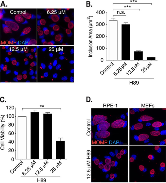
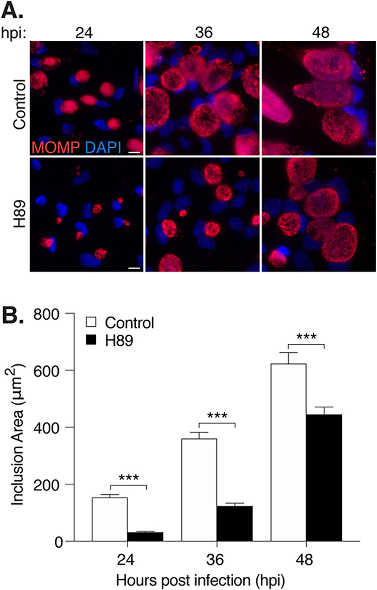
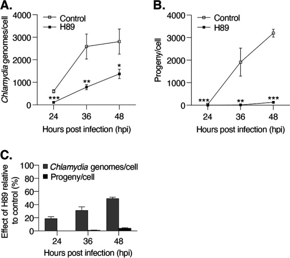
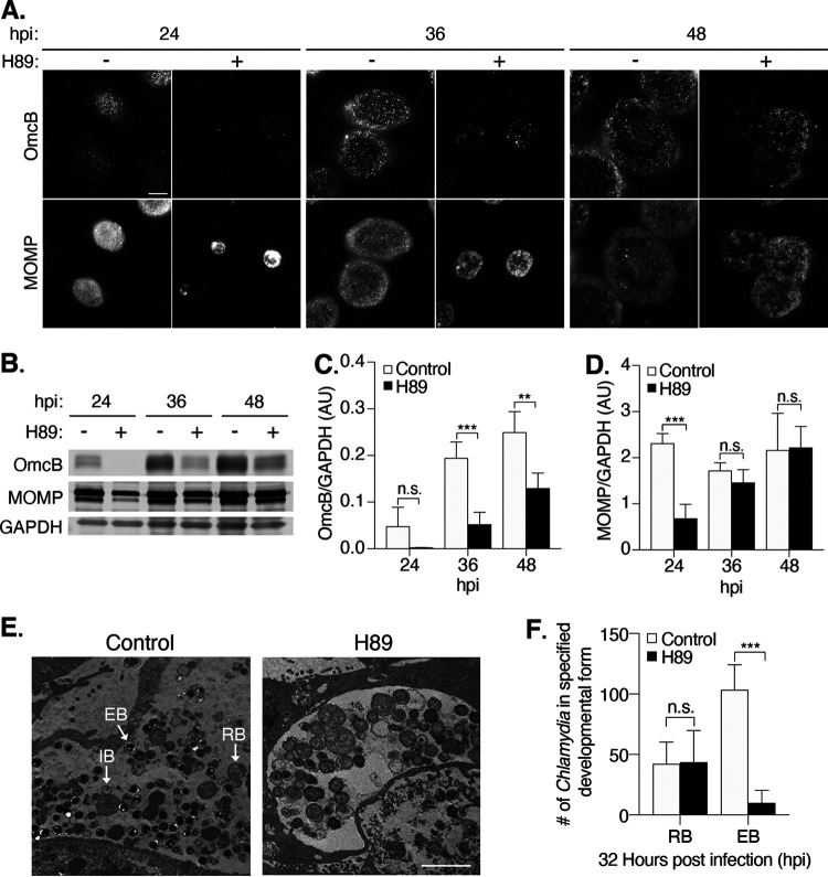
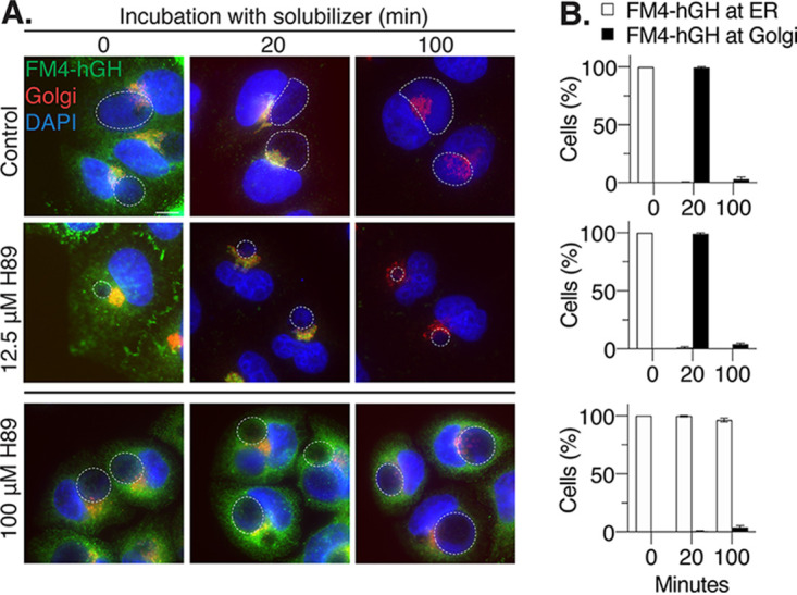
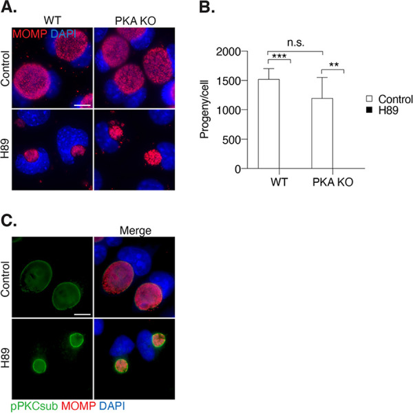
Chlamydia is an obligate intracellular bacterium and the most common reportable cause of human infection in the United States. This pathogen proliferates inside a eukaryotic host cell, where it resides within a membrane-bound compartment called the chlamydial inclusion. It has an unusual developmental cycle, marked by conversion between a replicating form, the reticulate body (RB), and an infectious form, the elementary body (EB). We found that the small molecule H89 slowed inclusion growth and decreased overall RB replication by 2-fold but caused a 25-fold reduction in infectious EBs. This disproportionate effect on EB production was mainly due to a defect in RB-to-EB conversion and not to the induction of chlamydial persistence, which is an altered growth state. Although H89 is a known inhibitor of specific protein kinases and vesicular transport to and from the Golgi apparatus, it did not cause these anti-chlamydial effects by blocking protein kinase A or C or by inhibiting protein or lipid transport. Thus, H89 is a novel anti-chlamydial compound that has a unique combination of effects on an intracellular Chlamydia infection.

Keywords: RB-to-EB conversion; developmental cycle; intracellular infection; isoquinoline sulfonamide.


Figures

FIG 1
FIG 1
H89 decreases C. trachomatis inclusion size. (A) HeLa cells grown on coverslips were infected with C. trachomatis L2 at an MOI of 3 and treated from 1 to 32 h postinfection (hpi) with the indicated concentrations of H89. Control cells (control) were incubated in the equivalent volume of DMSO, which served as the solvent for H89. In these immunofluorescence images, chlamydiae are detected with antibodies to the major out membrane protein MOMP (red) and chlamydial and host DNA are stained with DAPI (blue). Scale bar, 10 μm. (B) Quantification of the inclusion areas of the cells in panel A. One hundred inclusions were measured for each condition. Data from one representative experiment (n = 3) are shown as means ± SD; ***, P < 0.001. n.s. indicates the data are not statistically significant. (C) Viability of uninfected HeLa cells incubated with different concentrations of H89 for 32 h was assessed with an MTT cell viability assay. Data were normalized to the viability of untreated cells and are expressed as a percentage. Data are presented as means ± SD (n = 3); **, P ≤ 0.01. (D) Immunofluorescence images of retinal pigment epithelial cells (RPE-1) and mouse embryonic fibroblasts (MEFs) grown on coverslips, infected with C. trachomatis L2 at an MOI of 3, and treated with 12.5 μM H89 from 1 to 32 hpi. Scale bar, 10 μm.
FIG 2
FIG 2
H89 delays Chlamydia inclusion growth. (A) Immunofluorescence images of Chlamydia trachomatis L2-infected HeLa cells treated with H89 starting at 1 hpi. Samples were fixed at the indicated time points. Chlamydiae were stained with antibodies to MOMP (red), while chlamydial and host DNA was detected with DAPI (blue). Scale bar is 10 μm. (B) Quantification of inclusion surface areas for the cells in panel A. One hundred inclusions were measured for each condition. Data from one representative experiment are presented as means ± SD (n = 3); ***, P < 0.001.
FIG 3
FIG 3
H89 causes a greater effect on infectious progeny than on chlamydial replication. (A) The number of chlamydial genomes in infected, H89-treated HeLa cells was determined via qPCR at the indicated time points and normalized to the number of host cells. Data are presented as means ± SD (n = 3). **, P ≤ 0.01; ***, P ≤ 0.001. (B) The number of infectious EBs was determined in a progeny assay for infected control and H89-treated HeLa cells at the indicated time points and normalized to the number of host cells. Data are presented as means ± SD (n = 3); **, P ≤ 0.01; ***, P < 0.001. (C) The numbers of chlamydial genomes and infectious progeny in H89-treated samples were normalized to their untreated control counterparts (control = 100%) and expressed as a percentage to indicate relative effect of H89.
FIG 4
FIG 4
H89 delays expression of an EB-specific protein and reduces the number of EBs. (A) C. trachomatis L2-infected HeLa cells were treated with H89 at 1 hpi and imaged at the indicated time points. EBs were stained with antibodies to OmcB, while all chlamydial developmental forms were detected with antibodies to MOMP. Scale bar, 10 μm. (B) Western blot analysis of total cell lysates from infected cells treated as described for panel A. The levels of OmcB and MOMP are shown. GAPDH served as a loading control. A representative blot is shown (n = 4). (C) Quantification of OmcB protein levels from the Western blots in panel B. OmcB levels were normalized to the GAPDH loading control. The data are presented as arbitrary units (AU) and means ± SD (n = 4); **, P ≤ 0.01; ***, P ≤ 0.001. n.s. indicates that the data are not statistically significant. (D) Quantification of MOMP protein levels from the Western blots in panel B. MOMP levels were normalized to the GAPDH loading control. Data are presented as arbitrary units (AU; mean ± SD) (n = 4); ***, P ≤ 0.001. n.s. indicates the data are not statistically significant. (E) Electron micrographs (EM) of infected cells untreated or treated with H89 from 1 to 32 hpi. Scale bar, 2 μm. The different chlamydial developmental forms are indicated: EB, elementary body; IB, intermediate body; and RB, reticulate body. (F) The numbers of EBs and RBs, detected by EM in 32-hpi inclusions of control and H89-treated infections, were quantified. The data are presented as means ± SD for control (n = 7 inclusions) and H89 (n = 8 inclusions) treatments; ***, P ≤ 0.001; n.s., not statistically significant.
FIG 5
FIG 5
H89 at 12.5 μM does not alter ER-to-Golgi or post-Golgi apparatus trafficking in Chlamydia-infected cells. (A) C1 HeLa cells, which express the GFP-tagged transport reporter protein FM4-hGH (green), were infected and treated with H89 at 1 hpi. At 32 hpi, cells were incubated with solubilizer for 0, 20, or 100 min and then fixed and processed for fluorescence microscopy analysis. The Golgi apparatus was visualized with antibodies to GM130 (red), and chlamydial and host DNA were stained with DAPI (blue). The bottom panel shows a control sample in which infected C1 HeLa cells were treated with 100 μM H89 from 30 to 32 hpi before the addition of solubilizer at 32 hpi. Chlamydial inclusions are outlined with white, dashed lines. Scale bar, 10 μm. (B) The number of cells with GFP-tagged FM4-hGH at either the ER or the Golgi apparatus was normalized to the control and expressed as a percentage for the indicated time points after solubilizer addition (minutes). The data are presented as means ± SD (n = 3).
FIG 6
FIG 6
H89 effects on Chlamydia development are not due to inhibition of PKA or PKC. (A) mpkCCD WT and PKA knockout (PKA KO) cell lines were infected with C. trachomatis L2 and either left untreated or treated with 12.5 μM H89 from 1 to 32 hpi. Cells were fixed and subjected to immunofluorescence microscopy analysis with antibodies to MOMP (red). Chlamydial and host DNA were stained with DAPI (blue). Scale bar, 10 μm. (B) Production of infectious EBs (progeny assay) was measured for the cells treated as described for panel A, normalized to the number of host cells, and presented as means ± SD (n = 3); **, P ≤ 0.01; ***, P < 0.001; n.s., not statistically significant. (C) Immunofluorescence images of C. trachomatis L2-infected HeLa cells fixed at 24 hpi and stained with an antibody that recognizes phosphorylated PKC substrates (green). Chlamydiae were detected with antibodies to MOMP (red). Chlamydial and host DNA were stained with DAPI (blue). Scale bar, 10 μm.

Similar articles

Cited by

References

    1. Centers for Disease Control and Prevention. 2019. CDC–Chlamydia statistics. Centers for Disease Control and Prevention, Atlanta, GA. https://www.cdc.gov/std/chlamydia/stats.htm.
    1. Batteiger BE, Tan M. 2019. Chlamydia trachomatis (trachoma and urogenital infections), p 2301–2319. In Principles and practice of infectious diseases. Elsevier Inc., Philadelphia, PA.
    1. Moulder JW. 1991. Interaction of chlamydiae and host cells in vitro. Microbiol Rev 55:143–190. 10.1128/MR.55.1.143-190.1991. - DOI - PMC - PubMed
    1. Lee JK, Enciso GA, Boassa D, Chander CN, Lou TH, Pairawan SS, Guo MC, Wan FYM, Ellisman MH, Sütterlin C, Tan M. 2018. Replication-dependent size reduction precedes differentiation in Chlamydia trachomatis. Nat Commun 9:45. 10.1038/s41467-017-02432-0. - DOI - PMC - PubMed
    1. Snavely EA, Kokes M, Dunn JD, Saka HA, Nguyen BD, Bastidas RJ, McCafferty DG, Valdivia RH. 2014. Reassessing the role of the secreted protease CPAF in Chlamydia trachomatis infection through genetic approaches. Pathog Dis 71:336–351. 10.1111/2049-632X.12179. - DOI - PMC - PubMed

Publication types

MeSH terms

Substances