. 2021 Aug 16;89(9):e0005921.
doi: 10.1128/IAI.00059-21. Epub 2021 Aug 16.

Neonatal Enteropathogenic Escherichia coli Infection Disrupts Microbiota-Gut-Brain Axis Signaling

Affiliations

Neonatal Enteropathogenic Escherichia coli Infection Disrupts Microbiota-Gut-Brain Axis Signaling

Carly Hennessey et al. Infect Immun. .

Abstract

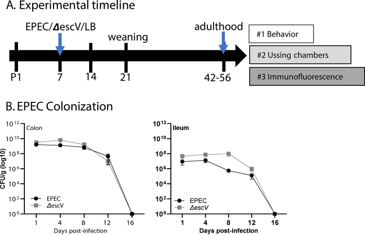
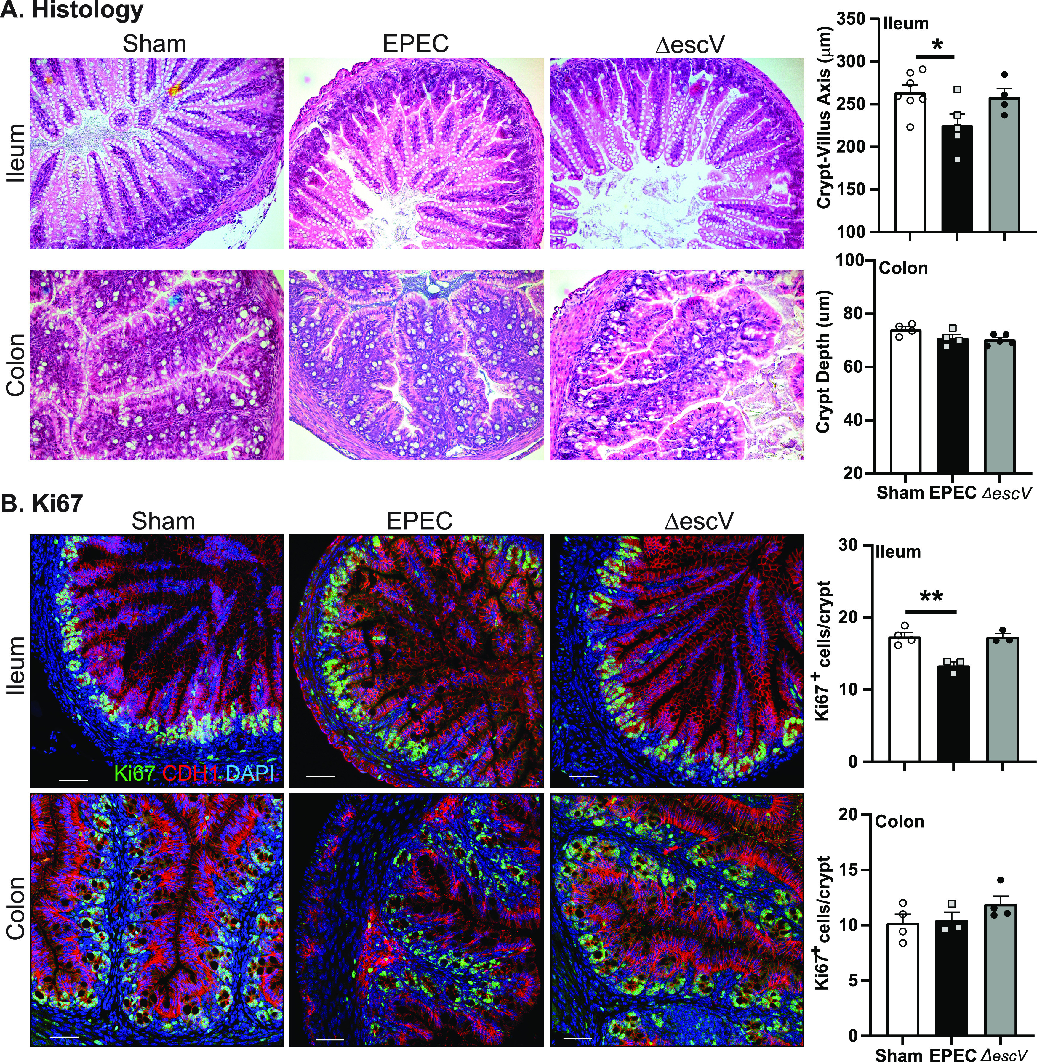
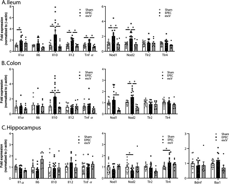
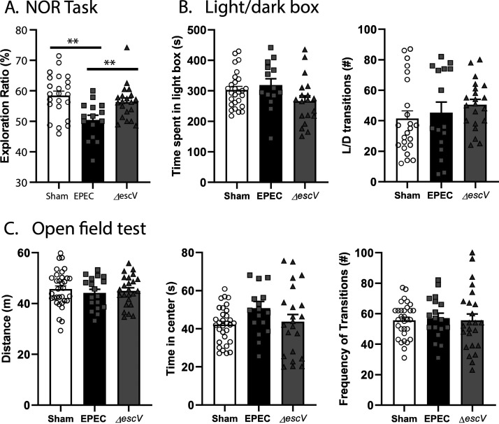
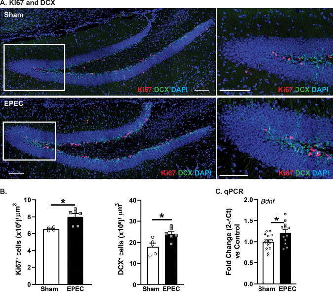
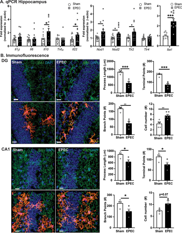
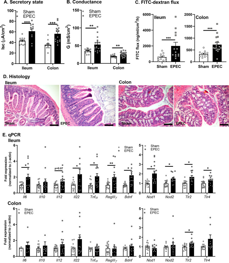
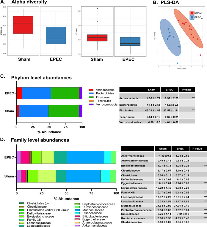
Diarrheal diseases are a leading cause of death in children under the age of 5 years worldwide. Repeated early-life exposures to diarrheal pathogens can result in comorbidities including stunted growth and cognitive deficits, suggesting an impairment in the microbiota-gut-brain (MGB) axis. Neonatal C57BL/6 mice were infected with enteropathogenic Escherichia coli (EPEC) (strain e2348/69; ΔescV [type III secretion system {T3SS} mutant]) or the vehicle (Luria-Bertani [LB] broth) via orogastric gavage at postnatal day 7 (P7). Behavior (novel-object recognition [NOR] task, light/dark [L/D] box, and open-field test [OFT]), intestinal physiology (Ussing chambers), and the gut microbiota (16S Illumina sequencing) were assessed in adulthood (6 to 8 weeks of age). Neonatal infection of mice with EPEC, but not the T3SS mutant, caused ileal inflammation in neonates and impaired recognition memory (NOR task) in adulthood. Cognitive impairments were coupled with increased neurogenesis (Ki67 and doublecortin immunostaining) and neuroinflammation (increased microglia activation [Iba1]) in adulthood. Intestinal pathophysiology in adult mice was characterized by increased secretory state (short-circuit current [Isc]) and permeability (conductance) (fluorescein isothiocyanate [FITC]-dextran flux) in the ileum and colon of neonatally EPEC-infected mice, along with increased expression of proinflammatory cytokines (Tnfα, Il12, and Il6) and pattern recognition receptors (Nod1/2 and Tlr2/4). Finally, neonatal EPEC infection caused significant dysbiosis of the gut microbiota, including decreased Firmicutes, in adulthood. Together, these findings demonstrate that infection in early life can significantly impair the MGB axis in adulthood.

Keywords: bacterial infection; behavior; microbiota-gut-brain axis; neurogenesis; neuroinflammation.


Figures

FIG 1
FIG 1
Study design and EPEC colonization. (A) Mouse pups were challenged at P7 (EPEC, ΔescV, or LB broth [sham]), and adults were divided into 3 experimental groups. (B) Colonic (left) and ileum (right) samples grown on MacConkey agar plates at 1, 4, 8, and 12 days postinfection to quantify EPEC and ΔescV colonization. Data represent 1 to 2 pups per litter from 3 to 4 separate litters per group per time point.
FIG 2
FIG 2
Neonatal GI histology. (A) Representative histological images (H&E staining) of ileum and colon from sham-, EPEC-, and ΔescV-challenged mice at 7 days postinfection (left) and quantification of the crypt-villus axis (ileum) and crypt depth (colon) (right). (B) Representative images of epithelial cell proliferation assessment by Ki67 (proliferation marker) (green), CDH1 (epithelial cell marker) (red), and DAPI (nuclei) (blue) in the ileum and colon (left) and quantification of cell numbers (right) (n = 3 to 5 mice per group) (*, P ≤ 0.05; **, P ≤ 0.01 [by one-way ANOVA and Tukey’s post hoc test]). Bars = 100 μm.
FIG 3
FIG 3
Neonatal GI inflammation. Relative mRNA expression levels in the ileum (A), colon (B), and hippocampus (C) at P14 are shown for cytokines and pattern recognition receptors (n = 8 to 16 mice per group) (*, P ≤ 0.05 [by one-way ANOVA and Tukey’s post hoc test]).
FIG 4
FIG 4
Neonatal EPEC infection induces cognitive deficits in adulthood. (A) Recognition memory was assessed using the novel-object recognition (NOR) task by quantification of the exploration ratio (percent). (B) Anxiety-like behavior was assessed using the light/dark (L/D) box by measuring the time spent in the light box and transitions between the light and dark boxes. (C) General locomotor activity was determined using the open-field test (OFT), by measuring the total distance traveled, frequency of transitions to the inner zone, and time spent in the inner zone (n = 15 to 34 mice per group) (***, P < 0.001 [by one-way ANOVA and Holm-Sidak’s post hoc test]).
FIG 5
FIG 5
Neonatal EPEC infection impairs hippocampal neurogenesis in adulthood. (A) Confocal imaging was used to assess neurogenesis via quantification of cell proliferation (Ki67) (red) and immature neurons (doublecortin [DCX]) (green). Bars = 100 μm. (B) Quantification of Ki67+ and DCX+ cells per cubic micrometer in the dentate gyrus (DG) (n = 5 mice per group) (*, P ≤ 0.05 [by Student’s t test]). (C) Relative mRNA expression of Bdnf in the hippocampus (n = 16 mice per group) (*, P ≤ 0.05 [by Student’s t test]).
FIG 6
FIG 6
Neonatal EPEC infection increases hippocampal neuroinflammation in adulthood. (A) Relative mRNA expression in the hippocampus (n = 12 to 16 mice per group) (*, P ≤ 0.05; **, P ≤ 0.01 [by Student’s t test, with a Mann-Whitney test and Welch’s correction where appropriate]). (B) Confocal imaging and morphological characterization of microglia in the dentate gyrus (DG) (top) and CA1 region (bottom) of the hippocampus. (Left) Microglia (Iba1) (green) with single-cell overlays (red) identified with 3DMorph and morphology characterized using Imaris. (Right) Process lengths, terminal points, branch points, and cell numbers were quantified. Nuclei were stained with DAPI (n = 5 to 6 mice per group) (**, P < 0.01; ***, P < 0.001 [by Student’s t test]). Bars = 15 μm.
FIG 7
FIG 7
Neonatal EPEC infection caused impaired intestinal physiology in adulthood. (A to C) Ussing chambers were used to assess the secretory state (short-circuit current [Isc]) (A) and tight junction permeability via conductance (G) (B) and FITC-dextran flux (C) in both the ileum and colon. (D) Histology of ileum (left) and colon (right) with representative H&E staining. (E) Relative mRNA expression in the ileum and colon (n = 12 to 16 mice per group) (*, P ≤ 0.05; **, P ≤ 0.01; ***, P < 0.001 [by Student’s t test, with a Mann-Whitney test and Welch’s correction where appropriate]).
FIG 8
FIG 8
Dysbiosis in adult mice following neonatal EPEC infection. 16S rRNA Illumina sequencing was performed on fecal pellets to characterize the microbiota. (A) Shannon and Chao1 measures of alpha diversity. (B) Multivariate analysis using partial least-squares discriminant analysis (PLS-DA) of genus-level abundances. (C and D) Phylum-level (C) and family-level (D) abundances, with the highest-abundance species presented and statistics outlined in the tables (n = 12 mice per group) (***, P < 0.001 [by a Holm-Sidak test]).

References

    1. Lanata CF, Fischer-Walker CL, Olascoaga AC, Torres CX, Aryee MJ, Black RE, Child Health Epidemiology Reference Group of the World Health Organization and UNICEF. 2013. Global causes of diarrheal disease mortality in children <5 years of age: a systematic review. PLoS One 8:e72788. 10.1371/journal.pone.0072788. - DOI - PMC - PubMed
    1. Donowitz M, Alpers DH, Binder HJ, Brewer T, Carrington J, Grey MJ. 2012. Translational approaches for pharmacotherapy development for acute diarrhea. Gastroenterology 142:e1–e9. 10.1053/j.gastro.2012.01.014. - DOI - PMC - PubMed
    1. Oria RB, Murray-Kolb LE, Scharf RJ, Pendergast LL, Lang DR, Kolling GL, Guerrant RL. 2016. Early-life enteric infections: relation between chronic systemic inflammation and poor cognition in children. Nutr Rev 74:374–386. 10.1093/nutrit/nuw008. - DOI - PMC - PubMed
    1. Niehaus MD, Moore SR, Patrick PD, Derr LL, Lorntz B, Lima AA, Guerrant RL. 2002. Early childhood diarrhea is associated with diminished cognitive function 4 to 7 years later in children in a northeast Brazilian shantytown. Am J Trop Med Hyg 66:590–593. 10.4269/ajtmh.2002.66.590. - DOI - PubMed
    1. MAL-ED Network Investigators. 2014. The MAL-ED study: a multinational and multidisciplinary approach to understand the relationship between enteric pathogens, malnutrition, gut physiology, physical growth, cognitive development, and immune responses in infants and children up to 2 years of age in resource-poor environments. Clin Infect Dis 59(Suppl 4):S193–S206. 10.1093/cid/ciu653. - DOI - PubMed

Publication types