. 2021 May 12;13(593):eabf1906.
doi: 10.1126/scitranslmed.abf1906. Epub 2021 Apr 5.

The neutralizing antibody, LY-CoV555, protects against SARS-CoV-2 infection in nonhuman primates

Affiliations

The neutralizing antibody, LY-CoV555, protects against SARS-CoV-2 infection in nonhuman primates

Bryan E Jones et al. Sci Transl Med. .

Abstract

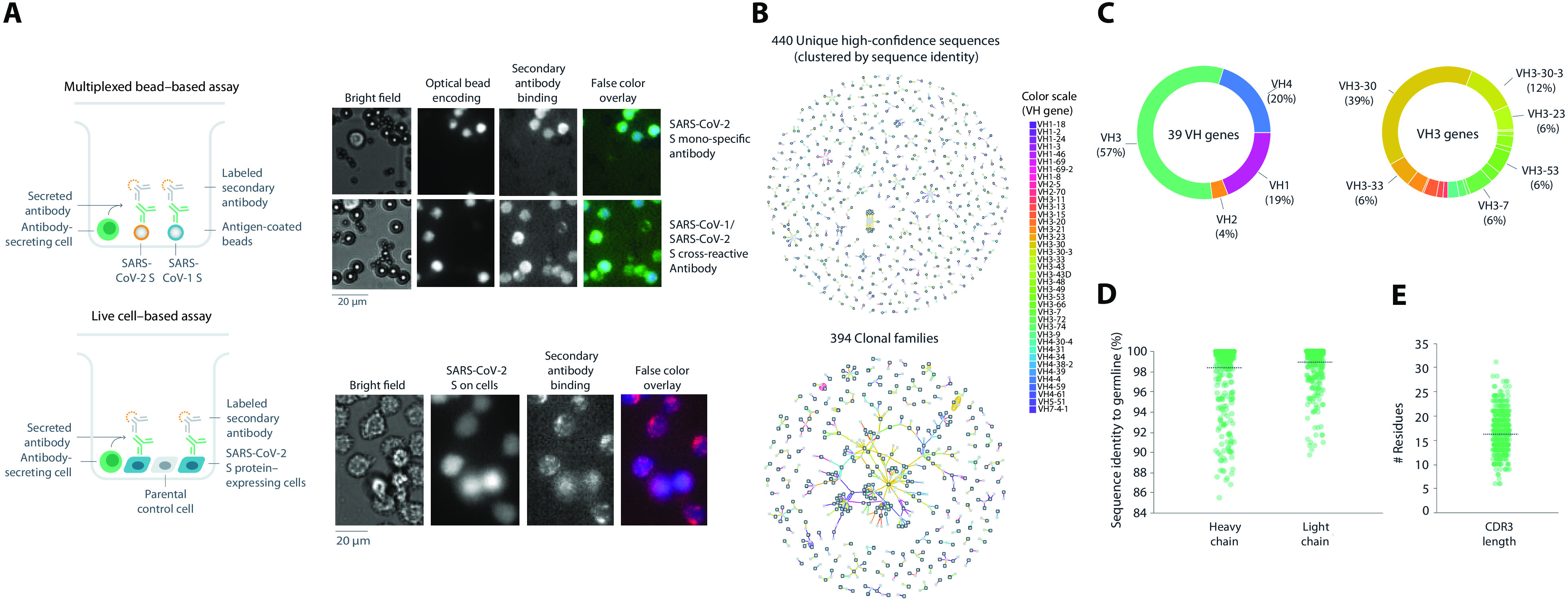
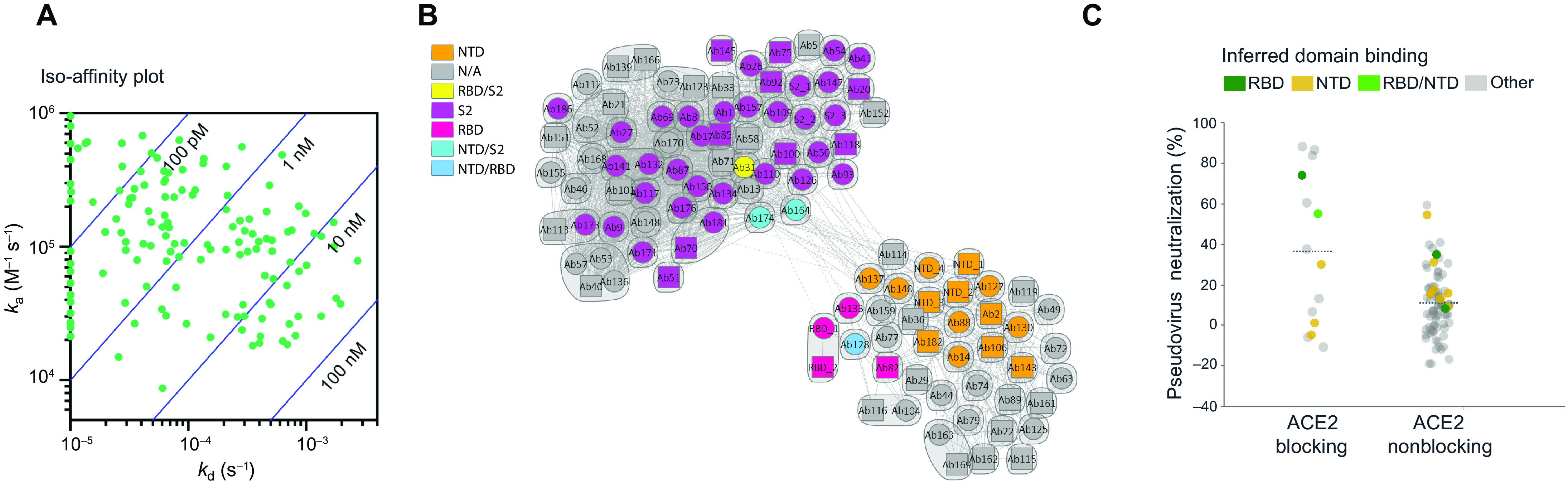
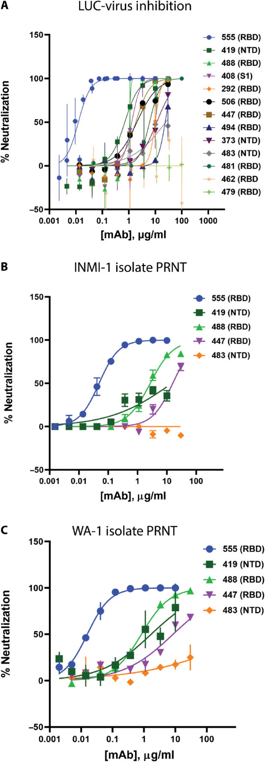
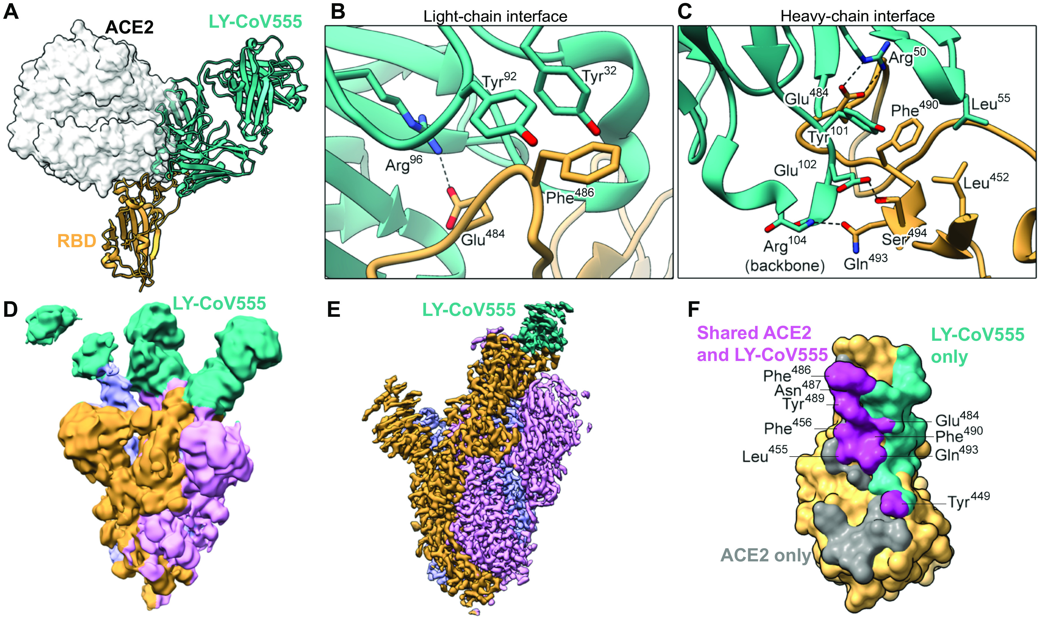
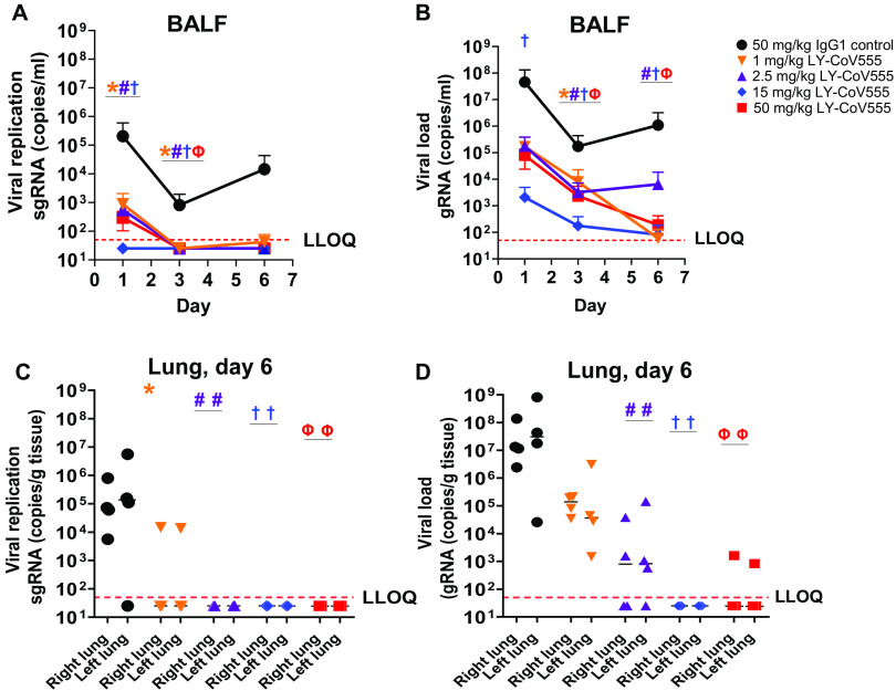
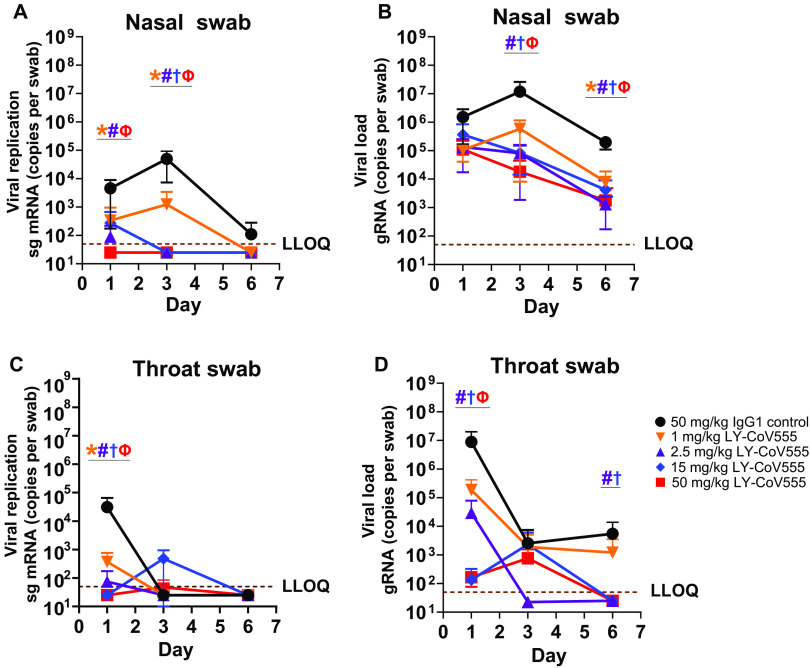
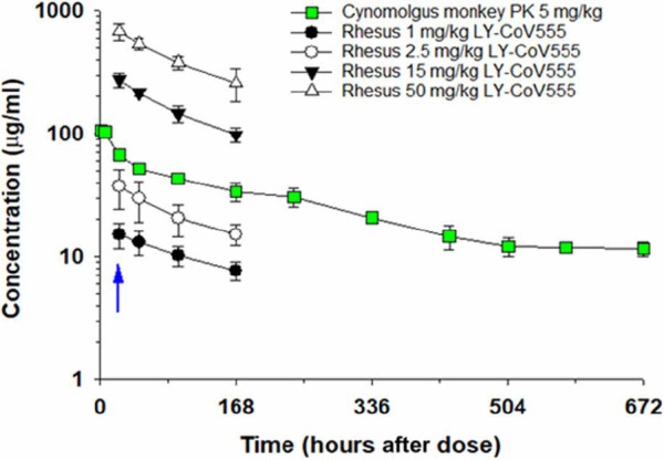
Severe acute respiratory syndrome coronavirus-2 (SARS-CoV-2) poses a public health threat for which preventive and therapeutic agents are urgently needed. Neutralizing antibodies are a key class of therapeutics that may bridge widespread vaccination campaigns and offer a treatment solution in populations less responsive to vaccination. Here, we report that high-throughput microfluidic screening of antigen-specific B cells led to the identification of LY-CoV555 (also known as bamlanivimab), a potent anti-spike neutralizing antibody from a hospitalized, convalescent patient with coronavirus disease 2019 (COVID-19). Biochemical, structural, and functional characterization of LY-CoV555 revealed high-affinity binding to the receptor-binding domain, angiotensin-converting enzyme 2 binding inhibition, and potent neutralizing activity. A pharmacokinetic study of LY-CoV555 conducted in cynomolgus monkeys demonstrated a mean half-life of 13 days and a clearance of 0.22 ml hour-1 kg-1, consistent with a typical human therapeutic antibody. In a rhesus macaque challenge model, prophylactic doses as low as 2.5 mg/kg reduced viral replication in the upper and lower respiratory tract in samples collected through study day 6 after viral inoculation. This antibody has entered clinical testing and is being evaluated across a spectrum of COVID-19 indications, including prevention and treatment.


Figures

Fig. 1
Fig. 1. Antibody screening and sequence analysis.
(A) Representation of multiplexed bead–based and live cell–based screening assays. Representative microscopic images of antibodies assessed for SARS-CoV-2 spike protein specificity in each indicated assay. (B) Sequence analysis of the 440 unique high-confidence paired-chain antibodies. Graphical representation of antibodies clustered according to sequence identity (top) or clonal family relationships (bottom). Each node indicates a chain or a cluster of chains. Heavy chains are outlined in black. Each line connecting the nodes indicates a single antibody, colored by VH gene usage according to legend. Multiple lines that connect to the same heavy- and light-chain clusters represent clonally related antibodies. (C to E) Sequence profiles of antibodies showing VH gene usage (C), distributions of sequence identity to germline for heavy and light chains (D), and CDR3 length (E). CDR3, complementary-determining region 3; VH, heavy chain variable.
Fig. 2
Fig. 2. Binding validation, kinetic analysis, and pseudovirus neutralization of SARS-CoV-2 spike protein–specific antibodies.
(A) Iso-affinity plot showing binding kinetics of recombinantly expressed antibodies. Association and dissociation rate constants were measured by high-throughput surface plasmon resonance (SPR) capture kinetic experiments with antibodies as immobilized ligands and antigens of interest as analytes. The distribution of kinetic values is displayed in an iso-affinity plot. Blue diagonal lines represent Kd values. ka, association kinetic rate constant; kd, dissociation kinetic rate constant; Kd, binding affinity constant. (B) Competition plot of recombinantly expressed antibodies. Each antibody was tested in two orientations: as a ligand on a chip and as an analyte in solution. Individual antibodies are represented either as a circle (data present in both orientations) or as a square (data present with the antibody in a single orientation). Bins are represented as envelopes (95 total) and competition between antibodies as solid (symmetric competition) or dashed (asymmetric competition) lines. Benchmark-based blocking profiles are indicated by color. N/A, not available; RBD, receptor-binding domain; NTD, N-terminal domain. (C) Pseudovirus neutralization activity relative to ACE2-blocking profile of antibodies. Where available, inferred binding domains of antibodies to RBD and NTD are indicated by color.
Fig. 3
Fig. 3. In vitro neutralization of SARS-CoV-2.
(A) Neutralization of recombinant SARS-CoV-2 encoding a nanoluciferase reporter in the Orf7a/b locus (Luc-Virus) in infected Vero E6 cells 24 hours after inoculation is shown. Values plotted are means of two replicates (n = 2), with error bars showing SEM. (B and C) Results from plaque reduction neutralization test (PRNT) assays for INMI-1 isolate (B) and 2020/USA/WA-1 isolate (C) of SARS-CoV-2 in Vero E6 cells 72 hours after inoculation are shown. Values plotted are means of two replicates (n = 2), with error bars showing SEM. mAb, monoclonal antibody; RBD, receptor-binding domain; NTD, N-terminal domain.
Fig. 4
Fig. 4. LY-CoV555 blocks ACE2 and binds to the spike protein RBD in up and down conformations.
(A) Crystal structure of the RBD-LY-CoV555 complex superimposed with the ACE2 receptor from a structure of the RBD-ACE2 complex (Protein Data Bank ID: 6M0J) (83). (B and C) Zoomed-in view of key atomic interactions at the interface of the LY-CoV555 light chain (B) and heavy chain (C) with the spike RBD. (D) Cryo-EM structure of the LY-CoV555 spike complex low-pass–filtered to 8-Å resolution and shown at low threshold to visualize all three Fabs (shown in cyan). (E) High-resolution cryo-EM map of the LY-CoV555-spike complex. (F) SARS-CoV-2 RBD molecular surface, with the portion of the surface that only interacts with ACE2 (gray), only interacts with LY-CoV555 (cyan), or interacts with both ACE2 and LY-CoV555 (pink). Interacting atoms were defined as being within 5.5 Å of each other, and the residues containing atoms interacting with both ACE2 and LY-CoV555 are labeled. Cryo-EM, cryo–electron microscopy; RBD, receptor-binding domain.
Fig. 5
Fig. 5. LY-CoV555 pretreatment reduces viral replication and load in the lower respiratory tract of rhesus macaques challenged with SARS-CoV-2.
Rhesus macaques (n = 3 or 4 per group) received LY-CoV555 (1, 2.5, 15, or 50 mg/kg) as a single intravenous dose 24 hours before SARS-CoV-2 inoculation. (A) sgRNA (viral replication) and (B) gRNA (viral load) were assessed by qRT-PCR in BALF over the course of 6 days after inoculation. (C) sgRNA (viral replication) and (D) gRNA (viral load) were assessed by qRT-PCR in lung tissue on day 6. Values represent the mean and SEM for three or four animals (A to C) or the mean of three or four animals (D). Samples below the lower limit of quantification (LLOQ) were designated a value of ½ LLOQ for plotting. LLOQ = 50 copies for genomic or subgenomic mRNA. Statistical testing results comparing treatment to the corresponding IgG1 control are provided in table S6. * denotes q value < 0.05, 1 mg/kg; # denotes q value < 0.05, 2.5 mg/kg; † denotes q value < 0.05, 15 mg/kg; and Ф denotes q value < 0.05, 50 mg/kg. BALF, bronchoalveolar lavage; gRNA, genomic RNA; qRT-PCR, quantitative real-time polymerase chain reaction; sgRNA, subgenomic RNA.
Fig. 6
Fig. 6. LY-CoV555 pretreatment reduces viral replication and load in the upper respiratory tract of rhesus macaques challenged with SARS-CoV-2.
Rhesus macaques (n = 3 or 4 per group) received LY-CoV555 (1, 2.5, 15, or 50 mg/kg) as a single intravenous dose 24 hours before viral challenge. (A) sgRNA (viral replication) and (B) gRNA (viral load) were assessed by qRT-PCR in nasal swabs over 6 days after SARS-CoV-2 inoculation. (C) sgRNA (viral replication) and (D) gRNA (viral load) were assessed by qRT-PCR in throat swabs over 6 days after SARS-CoV-2 inoculation. Values represent the mean ± SEM for three or four animals at indicated time points. Samples below the lower limit of quantification (LLOQ) were designated a value of ½ LLOQ for plotting. LLOQ = 50 copies for gRNA or sgRNA. Statistical testing results comparing treatment to the corresponding IgG1 control are provided in table S6. * denotes q value < 0.05, 1 mg/kg; # denotes q value < 0.05, 2.5 mg/kg; † denotes q value < 0.05, 15 mg/kg; and Ф denotes q value < 0.05, 50 mg/kg. gRNA, genomic RNA; qRT-PCR, quantitative real-time polymerase chain reaction; sg mRNA, subgenomic RNA.
Fig. 7
Fig. 7. Determination of serum concentrations of LY-CoV555 in a cynomolgus monkey PK study and in rhesus macaques during SARS-CoV-2 infection.
In the PK study, female cynomolgus monkeys received LY-CoV555 (5 mg/kg) as a single intravenous dose, and serum samples were collected through 672 hours after administration. Samples were analyzed using a human IgG ELISA or a ligand-capture LC-MS assay, which provided comparable results. Data points represent mean ± SD of determination from three animals. Cynomolgus monkey PK data are represented by green squares. In the rhesus macaque challenge experiments, animals (n = 3 or 4 per group) were administered LY-CoV555 (1, 2.5, 15, or 50 mg/kg) as a single intravenous dose, and serum samples were collected on study day −1 (predose) and days 0, 1, 3, and 6 (24, 48, 96, and 168 hours after intravenous dosing). Data points represent the mean ± SD for three or four animals. The blue arrow refers to viral challenge in rhesus macaque study on study day 0 (24 hours after intravenous administration of LY-CoV555).

Update of

References

    1. McKee M., Stuckler D., If the world fails to protect the economy, COVID-19 will damage health not just now but also in the future. Nat. Med. 26, 640–642 (2020). - PubMed
    1. Huang C., Wang Y., Li X., Ren L., Zhao J., Hu Y., Zhang L., Fan G., Xu J., Gu X., Cheng Z., Yu T., Xia J., Wei Y., Wu W., Xie X., Yin W., Li H., Liu M., Xiao Y., Gao H., Guo L., Xie J., Wang G., Jiang R., Gao Z., Jin Q., Wang J., Cao B., Clinical features of patients infected with 2019 novel coronavirus in Wuhan, China. Lancet 395, 497–506 (2020). - PMC - PubMed
    1. Jiang S., Hillyer C., Du L., Neutralizing antibodies against SARS-CoV-2 and other human coronaviruses. Trends Immunol. 41, 545 (2020). - PMC - PubMed
    1. Williamson E. J., Walker A. J., Bhaskaran K., Bacon S., Bates C., Morton C. E., Curtis H. J., Mehrkar A., Evans D., Inglesby P., Cockburn J., McDonald H. I., MacKenna B., Tomlinson L., Douglas I. J., Rentsch C. T., Mathur R., Wong A. Y. S., Grieve R., Harrison D., Forbes H., Schultze A., Croker R., Parry J., Hester F., Harper S., Perera R., Evans S. J. W., Smeeth L., Goldacre B., Factors associated with COVID-19-related death using OpenSAFELY. Nature 584, 430–436 (2020). - PMC - PubMed
    1. Gilchuk P., Bombardi R. G., Erasmus J. H., Tan Q., Nargi R., Soto C., Abbink P., Suscovich T. J., Durnell L. A., Khandhar A., Archer J., Liang J., Fouch M. E., Davidson E., Doranz B. J., Jones T., Larson E., Ertel S., Granger B., Fuerte-Stone J., Roy V., Broge T., Linnekin T. C., Linde C. H., Gorman M. J., Nkolola J., Alter G., Reed S. G., Barouch D. H., Diamond M. S., Crowe J. E. Jr., Van Hoeven N., Thackray L. B., Carnahan R. H., Integrated pipeline for the accelerated discovery of antiviral antibody therapeutics. Nat. Biomed. Eng. 4, 1030–1043 (2020). - PMC - PubMed

Publication types

Substances