. 2021 Apr 4;13(7):10490-10516.
doi: 10.18632/aging.202810. Epub 2021 Apr 4.

Nuclear envelope tethering inhibits the formation of ALT-associated PML bodies in ALT cells

Affiliations

Nuclear envelope tethering inhibits the formation of ALT-associated PML bodies in ALT cells

Chia-Wei Yang et al. Aging (Albany NY). .

Abstract

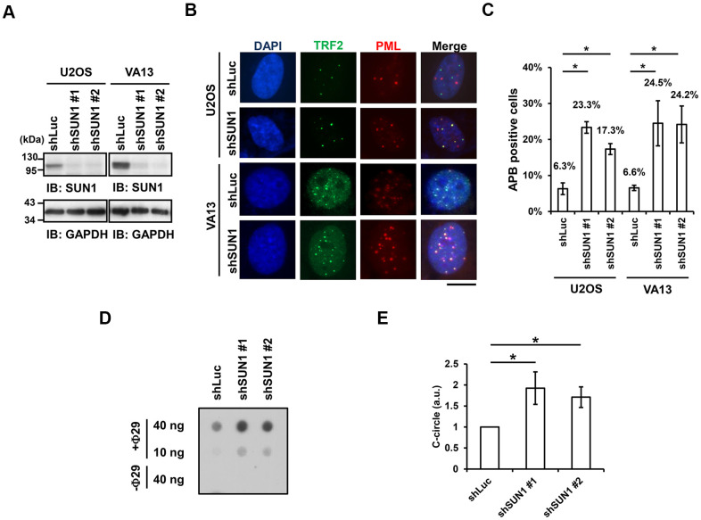
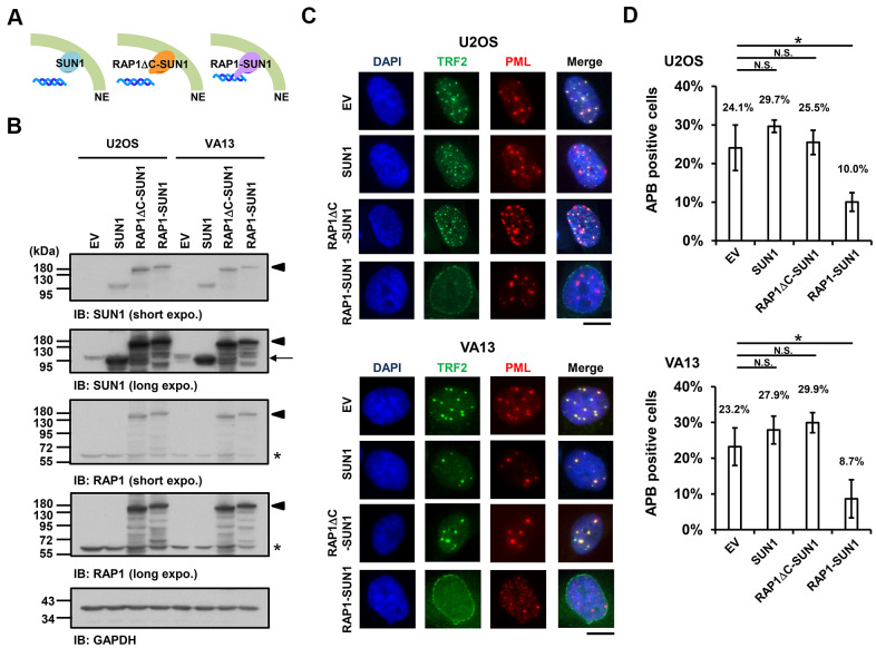
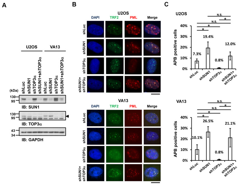
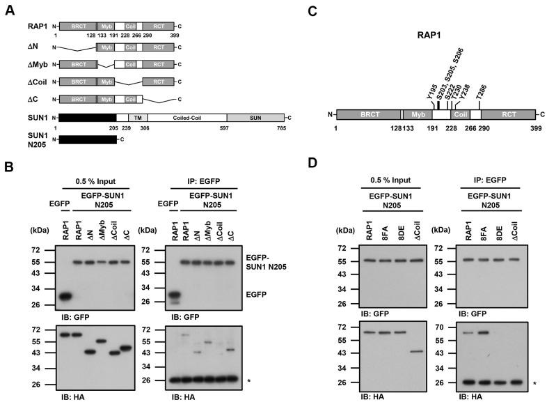
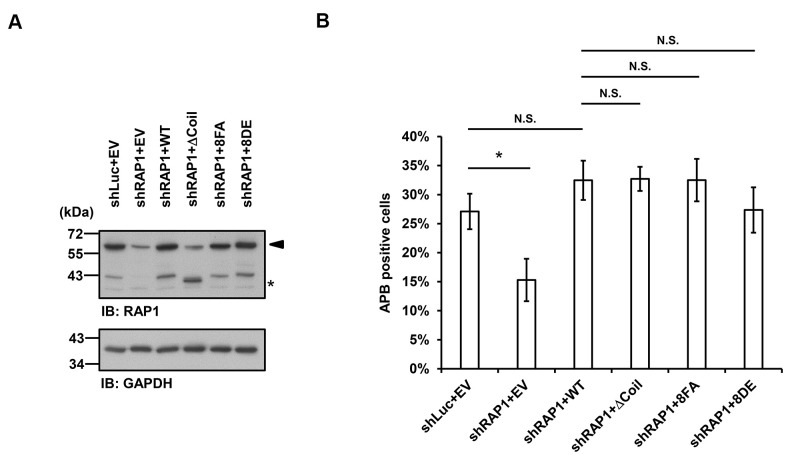
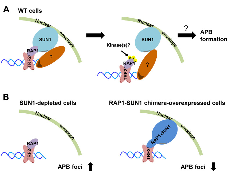
Telomere length homeostasis is essential for maintaining genomic stability and cancer proliferation. Telomerase-negative cancer cells undergo recombination-mediated alternative lengthening of telomeres. Telomeres associate with the nuclear envelope through the shelterin RAP1 and nuclear envelope SUN1 proteins. However, how the associations between telomeres and the nuclear envelope affect the progression of telomere recombination is not understood. Here, we show that telomere anchorage might inhibit telomere-telomere recombination. SUN1 depletion stimulates the formation of alternative lengthening of telomeres-associated promyelocytic leukemia bodies in ALT cells. In contrast, overexpression of a telomere-nuclear envelope-tethering chimera protein, RAP1-SUN1, suppresses APB formation. Moreover, inhibition of this nuclear envelope attachment alleviates the requirement of TOP3α for resolving the supercoiling pressure during telomere recombination. A coimmunoprecipitation assay revealed that the SUN1 N-terminal nucleoplasmic domain interacts with the RAP1 middle coil domain, and phosphorylation-mimetic mutations in RAP1 inhibit this interaction. However, abolishing the RAP1-SUN1 interaction does not hinder APB formation, which hints at the existence of another SUN1-dependent telomere anchorage pathway. In summary, our results reveal an inhibitory role of telomere-nuclear envelope association in telomere-telomere recombination and imply the presence of redundant pathways for the telomere-nuclear envelope association in ALT cells.

Keywords: RAP1; SUN1; alternative lengthening of telomeres; nuclear envelope tethering; telomere-telomere recombination.


Conflict of interest statement

CONFLICTS OF INTEREST: The authors declare that they have no conflicts of interest.

Figures

Figure 1
Figure 1
SUN1 knockdown induces APB formation and C-circle levels. (A) U2OS and VA13 cells were infected with control (shLuc) or shSUN1 lentivirus and selected with 1 μg/ml puromycin for 3 days. Cell lysates were subjected to immunoblot analysis with anti-SUN1 and anti-GAPDH antibodies. GAPDH was used as the loading control. (B) Representative images show the colocalization of TRF2 and PML in U2OS cells (upper panel) and VA13 cells (bottom panel). Virus-infected and puromycin-selected cells were subjected to immunofluorescence staining with anti-TRF2 and anti-PML antibodies. DNA was stained with DAPI. Cells containing at least three large TRF2 and PML colocalization foci (yellow) in the nucleus were counted as APB-positive cells. Scale bar, 20 μm. (C) Quantification of APBs (%) in the U2OS and VA13 cells shown in (B). Approximately 200-300 cells were analyzed for each independent experiment. Error bars denote SD; n=3 (independent experiments); *P<0.05 (two-tailed Student’s t-test). (D) Depletion of SUN1 stimulates the formation of C-circles in U2OS cells. (E) Quantification of the level of C-circles in the cells in (D). The signals were quantified with ImageJ software. The level of C-circles is represented in an arbitrary unit (a.u.). Error bars denote SD; n=3 (independent experiments); *P<0.05 (two-tailed Student’s t-test).
Figure 2
Figure 2
The enhancement of nuclear envelope anchorage inhibits APB formation. (A) Schematic diagrams of cells overexpressing SUN1, RAP1-RCT-domain-deleted-SUN1 (RAP1ΔC-SUN1), or RAP1-SUN1 fusion chimera protein are shown. NE, the nuclear envelope. (B) U2OS and VA13 cells were infected with lentivirus expressing the empty vector control (EV), SUN1, RAP1ΔC-SUN1, or RAP1-SUN1 fusion and then selected in medium containing G418 for 5 days. Cell lysates were analyzed by immunoblotting with anti-RAP1, anti-SUN1, and anti-GAPDH antibodies. The arrowhead indicates the RAP1-SUN1 fusion protein. The arrow indicates endogenous SUN1. The asterisk indicates endogenous RAP1. The ladders under the major protein band show possible products of protein degradation. GAPDH was used as the loading control. (C) Representative images show the colocalization of TRF2 and PML in U2OS cells (upper panel) and VA13 cells (bottom panel), as shown in Figure 1. Scale bar, 20 μm. (D) Quantification of APBs (%) in the U2OS and VA13 cells shown in (C). Approximately 200-300 cells were analyzed for each independent experiment. Error bars denote SD; n=3 (independent experiments); *P<0.05 (two-tailed Student’s t-test). N.S., no significance.
Figure 3
Figure 3
SUN1 depletion increases APB formation in TOP3α-knockdown cells. (A) U2OS and VA13 cells were infected with control (shLuc), shSUN1, shTOP3α, or shSUN1 combined with shTOP3α lentiviruses and selected for 3 days. Cell lysates were analyzed by immunoblotting with anti-SUN1, anti-TOP3α, and anti-GAPDH antibodies. The arrowhead indicates the location of the TOP3α protein. GAPDH was used as the loading control. (B) Representative images show the colocalization of TRF2 and PML in U2OS cells (upper panel) and VA13 cells (bottom panel), as shown in Figure 1. Scale bar, 20 μm. (C) Quantification of APBs (%) in the U2OS and VA13 cells shown in (B). Approximately 200-300 cells were analyzed for each independent experiment. Error bars denote SD; n=3 (independent experiments); *P<0.05 (two-tailed Student’s t-test). N.S., no significance.
Figure 4
Figure 4
The coil region of RAP1 and the potential phosphorylation of residues in that domain are likely critical for the SUN1 interaction. (A) Schematic representations of the N-terminal HA-tagged RAP1 constructs and the N-terminal EGFP-tagged SUN1 constructs. TM, transmembrane. (B) U2OS cells were transfected with HA-tagged RAP1 together with either EGFP or EGFP-tagged SUN1 N205. Forty-eight hours posttransfection, the cells were harvested for use in immunoprecipitation assays. EGFP-SUN1 N205 was immunoprecipitated with anti-GFP beads. Input and immunoprecipitated proteins (IPs) were analyzed by immunoblotting with anti-GFP and anti-HA antibodies. Asterisk (*), nonspecific band. (C) A schematic representation of the eight potential phosphorylation sites in the coil domain of RAP1. (D) U2OS cells were transfected with HA-tagged RAP1 WT, nonphosphorylatable (8FA), or phospho-mimetic (8DE) mutant together with EGFP-tagged SUN1 N205. Forty-eight hours posttransfection, the cells were harvested for use in immunoprecipitation assays. Input and immunoprecipitated proteins (IPs) were analyzed by immunoblotting with anti-GFP and anti-HA antibodies. Asterisk (*), nonspecific band.
Figure 5
Figure 5
Disruption of the interaction between RAP1 and SUN1 does not interfere with APB formation in ALT cells. (A) U2OS cells were infected with the knockdown control (shLuc) or shRAP1 lentivirus and simultaneously complemented with control (EV), wild-type RAP1 (WT), RAP1 coil deletion (ΔCoil), nonphosphorylatable (8FA), or phospho-mimetic (8DE) RAP1 mutant lentiviruses. Virus-infected cells were selected for 5 days and subjected to further methionine restriction for 3 days. Cell lysates were analyzed by immunoblotting with anti-RAP1 and anti-GAPDH antibodies. The arrowhead indicates the location of endogenous RAP1, and the multiple lower-molecular-weight bands are degraded RAP1. Asterisk (*), RAP1 coil deletion mutant. GAPDH was used as the loading control. (B) Quantification of APBs (%) in the U2OS cells shown in (A). Approximately 200-300 cells were analyzed for each independent experiment. Error bars denote SD; n=3 (independent experiments); *P<0.05 (two-tailed Student’s t-test). N.S., no significance.
Figure 6
Figure 6
Model depicting the role of RAP1-SUN1-mediated telomere-nuclear envelope attachment during telomere-telomere recombination in ALT cells. (A) The interaction between RAP1 and SUN1 contributes to telomere anchorage to the nuclear envelope. Unknown kinases might phosphorylate the coil domain of RAP1, inducing RAP1 release from SUN1. Additionally, SUN1 might connect with an unknown telomere-binding protein, and this interaction may provide another telomere-nuclear envelope tethering mechanism to constrain the telomere from freely roaming. The molecular mechanism of how the telomeres depart from the nuclear envelope to the APB remains a mystery. (B) The depletion of SUN1 leads to the release telomeres from the nuclear envelope anchorage and increases the APB formation. However, the RAP1-SUN1 chimera enforces anchorage and decreases the APB formation in ALT cells.

Similar articles

Cited by

References

    1. Blackburn EH, Epel ES, Lin J. Human telomere biology: A contributory and interactive factor in aging, disease risks, and protection. Science. 2015; 350:1193–98. 10.1126/science.aab3389 - DOI - PubMed
    1. de Lange T. Protection of mammalian telomeres. Oncogene. 2002; 21:532–40. 10.1038/sj.onc.1205080 - DOI - PubMed
    1. Greider CW, Blackburn EH. Identification of a specific telomere terminal transferase activity in Tetrahymena extracts. Cell. 1985; 43:405–13. 10.1016/0092-8674(85)90170-9 - DOI - PubMed
    1. Artandi SE, DePinho RA. A critical role for telomeres in suppressing and facilitating carcinogenesis. Curr Opin Genet Dev. 2000; 10:39–46. 10.1016/S0959-437X(99)00047-7 - DOI - PubMed
    1. Hetzer MW, Walther TC, Mattaj IW. Pushing the envelope: structure, function, and dynamics of the nuclear periphery. Annu Rev Cell Dev Biol. 2005; 21:347–80. 10.1146/annurev.cellbio.21.090704.151152 - DOI - PubMed

Publication types

MeSH terms

LinkOut - more resources