Review
. 2021 May;37(5):1449-1459.
doi: 10.1007/s00381-021-05148-1. Epub 2021 Apr 5.

MRI-based diagnosis and treatment of pediatric brain tumors: is tissue sample always needed?

Affiliations
Review

MRI-based diagnosis and treatment of pediatric brain tumors: is tissue sample always needed?

Jehuda Soleman et al. Childs Nerv Syst. 2021 May.

Abstract

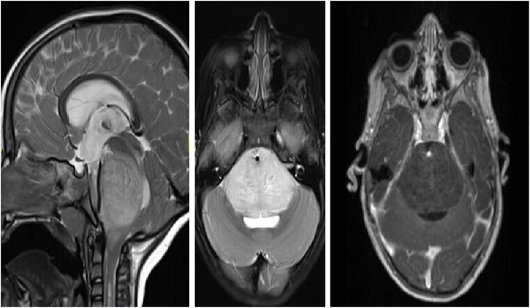
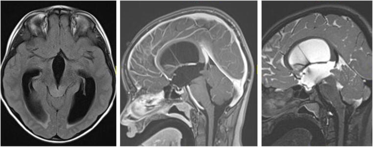
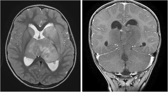
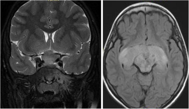
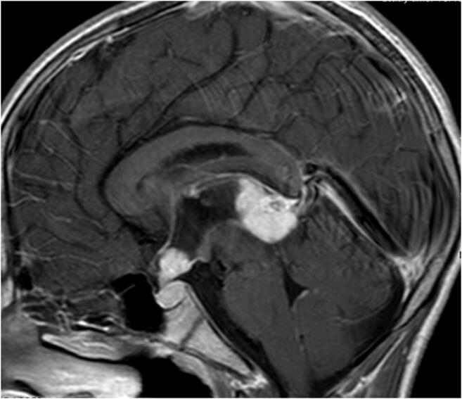
Traditional management of newly diagnosed pediatric brain tumors (PBTs) consists of cranial imaging, typically magnetic resonance imaging (MRI), and is frequently followed by tissue diagnosis, through either surgical biopsy or tumor resection. Therapy regimes are typically dependent on histological diagnosis. To date, many treatment regimens are based on molecular biology. The scope of this article is to discuss the role of diagnosis and further treatment of PBTs based solely on MRI features, in light of the latest treatment protocols. Typical MRI findings and indications for surgical biopsy of these lesions are described.

Keywords: Image-based diagnosis; Magnetic resonance imaging; Pediatric brain tumors; Pediatric neuroradiology; Pediatric neurosurgery; Surgical biopsy.


Conflict of interest statement

The authors report no conflict of interest concerning the materials or methods used in this study or the findings specified in this paper.

Figures

Fig. 1
Fig. 1
Diffuse intrinsic pontine glioma (DIPG): sagittal (left) and axial (middle) T2-weighted, and axial (right) T1-contrast-enhanced MRI of a 3.5-year-old girl presenting with new-onset cranial neuropathy, motor decline, and headaches. MRI shows a diffuse pontine tumor, with engulfment of the basilar artery, and very mild linear contrast enhancement. A shunt was placed to treat the hydrocephalus. Based on the MRI features, a DIPG was suspected and treated accordingly
Fig. 2
Fig. 2
Tectal plate glioma (TPG): axial FLAIR, and sagittal T2-weighted and T1-contrast-enhanced MRI of a 12-year-old girl presenting with obstructive hydrocephalus. MRI shows an isointense lesion on T1-weighted imaging (middle image), without contrast uptake, slightly hyperintense on T2-weighted imaging (right image), leading to a “ballooned” tectum and compression of the aqueduct. The child underwent an ETV and is symptom-free for last 6 years, with no tumor progression
Fig. 3
Fig. 3
Bilateral thalamic glioma (BTG): axial T2-weighted and coronal T1-contrast-enhanced MRI of a 4-year-old boy presenting with symptoms of increased intracranial pressure. MRI shows a diffuse bithalamic tumor, extending to the basal ganglia (especially on the left). An endoscopic third ventriculostomy and septostomy were performed. Patient succumbed to disease about 1 year later, following oncological treatment
Fig. 4
Fig. 4
Optic pathway glioma (OPG) in neurofibromatosis type I (NF1): coronal T2-weighted MRI and axial FLAIR MRI of a 9-year-old boy with NF1. MRI shows a typical OPG, involving the optic nerves, chiasm, and optic tracts. Typical NF changes are seen in the mid-brain. The child underwent treatment with vincristine and carboplatin, followed by vinblastine. Over the years, the tumor reduced in size; however, vision continued to deteriorate
Fig. 5
Fig. 5
Subependymal giant cell astrocytoma (SEGA): axial FLAIR and T1-contrast-enhanced MRI of a 12-year-old girl with known tuberous sclerosis and epilepsy, presenting with new-onset symptoms of increased intracranial pressure. MRI shows multiple cortical tubers, and bilateral SEGA (prominent on the right side) with obstructive hydrocephalus. The child underwent transcortical resection of the tumor and a septostomy. No progression of the left tumor or recurrence of the right tumor is evident over a 6-year follow-up
Fig. 6
Fig. 6
Bifocal (suprasellar/pineal) germ cell tumor (BGCT): sagittal T1-contrast-enhanced MRI of a 13-year-old boy presenting with obstructive hydrocephalus and diabetes insipidus. MRI shows a “bifocal–pineal and suprasellar” tumor. A shunt was placed. CSF and blood markers (AFP + BHCG) were negative. Patient was treated for a presumed germinoma and is tumor-free for more than 5 years. One year after shunt insertion, he underwent an endoscopic third ventriculostomy (ETV) due to a shunt malfunction

References

    1. Pierre-Aurélien B, Alexandru S, Federico DR, et al. Diffuse intrinsic pontine glioma in children: document or treat? World Neurosurg. 2016;93:485.e11–485.e14. doi: 10.1016/j.wneu.2016.07.011. - DOI - PubMed
    1. Cohen KJ, Jabado N, Grill J. Diffuse intrinsic pontine gliomas-current management and new biologic insights. Is there a glimmer of hope? Neuro Oncol. 2017;19:1025–1034. doi: 10.1093/neuonc/nox021. - DOI - PMC - PubMed
    1. Puget S, Beccaria K, Blauwblomme T, Roujeau T, James S, Grill J, Zerah M, Varlet P, Sainte-Rose C. Biopsy in a series of 130 pediatric diffuse intrinsic Pontine gliomas. Childs Nerv Syst. 2015;31:1773–1780. doi: 10.1007/s00381-015-2832-1. - DOI - PubMed
    1. Makepeace L, Scoggins M, Mitrea B, Li Y, Edwards A, Tinkle CL, Hwang S, Gajjar A, Patay Z. MRI patterns of extrapontine lesion extension in diffuse intrinsic pontine gliomas. Am J Neuroradiol. 2020;41:323–330. doi: 10.3174/ajnr.A6391. - DOI - PMC - PubMed
    1. Tisnado J, Young R, Peck KK, Haque S. Conventional and advanced imaging of diffuse intrinsic pontine glioma. J Child Neurol. 2016;31:1386–1393. doi: 10.1177/0883073816634855. - DOI - PMC - PubMed

LinkOut - more resources