. 2021 Oct 7;15(10):1737-1750.
doi: 10.1093/ecco-jcc/jjab061.

Potential Role of Epithelial Endoplasmic Reticulum Stress and Anterior Gradient Protein 2 Homologue in Crohn's Disease Fibrosis

Affiliations

Potential Role of Epithelial Endoplasmic Reticulum Stress and Anterior Gradient Protein 2 Homologue in Crohn's Disease Fibrosis

Sophie Vieujean et al. J Crohns Colitis. .

Abstract

Background and aims: Intestinal fibrosis is a common complication of Crohn's disease [CD]. It is characterised by an accumulation of fibroblasts differentiating into myofibroblasts secreting excessive extracellular matrix. The potential role of the intestinal epithelium in this fibrotic process remains poorly defined.

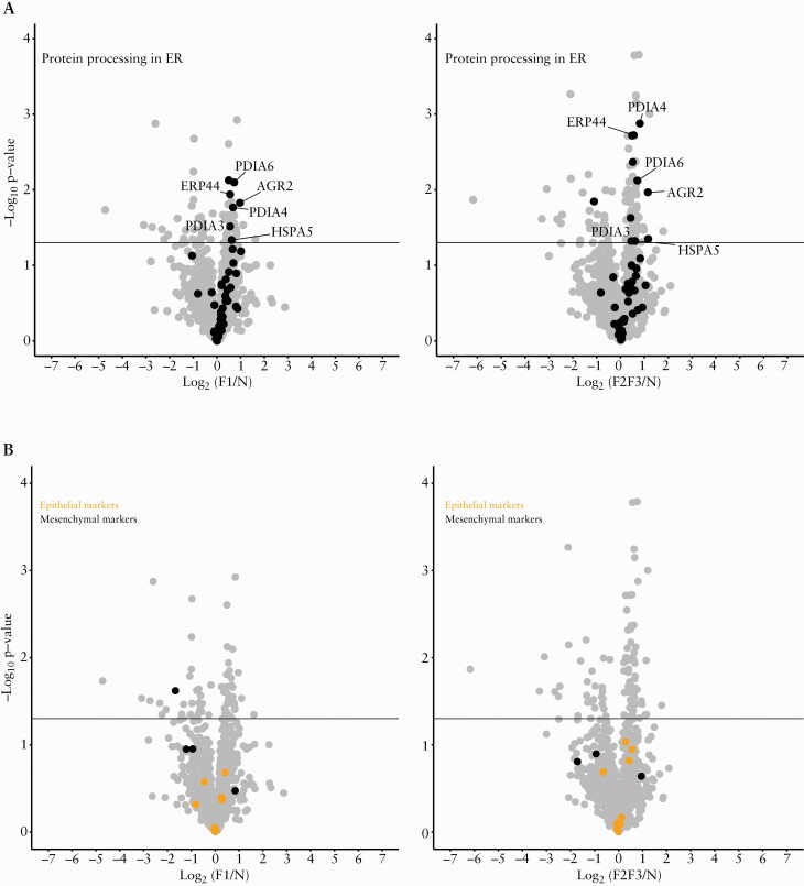
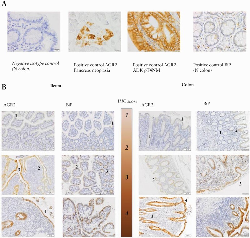
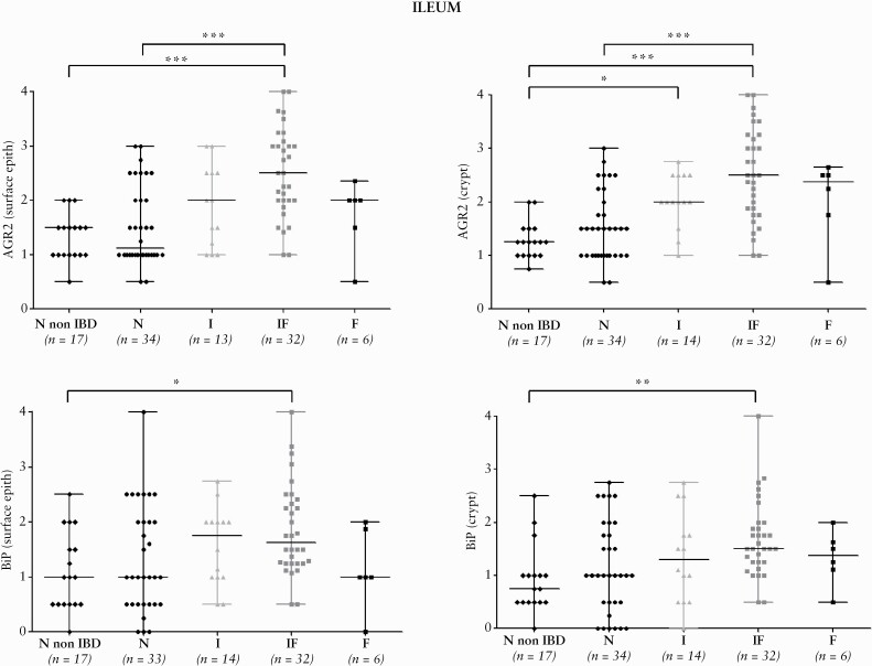
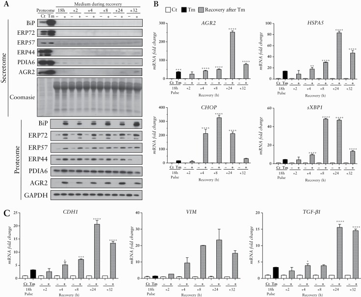
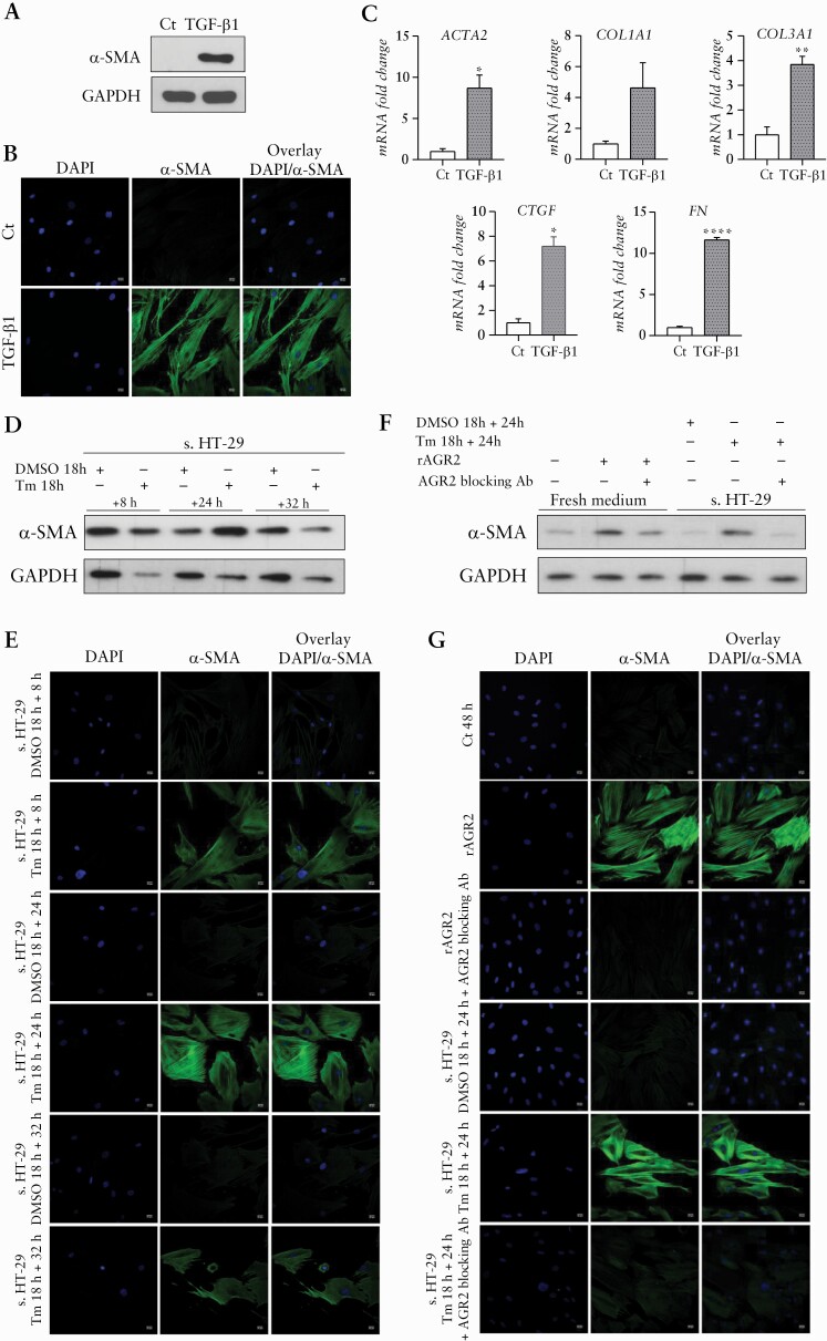
Methods: We performed a pilot proteomic study comparing the proteome of surface epithelium, isolated by laser-capture microdissection, in normal and fibrotic zones of resected ileal CD strictures [13 zones collected in five patients]. Proteins of interests were validated by immunohistochemistry [IHC] in ileal and colonic samples of stricturing CD [n = 44], pure inflammatory CD [n = 29], and control [n = 40] subjects. The pro-fibrotic role of one selected epithelial protein was investigated through in-vitro experiments using HT-29 epithelial cells and a CCD-18Co fibroblast to myofibroblast differentiation model.

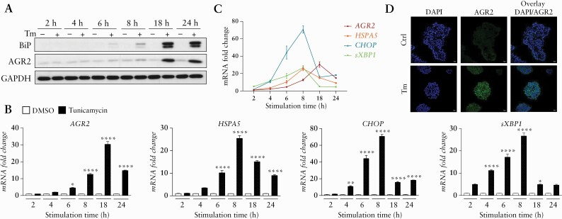
Results: Proteomic study revealed an endoplasmic reticulum [ER] stress proteins increase in the epithelium of CD ileal fibrotic strictures, including anterior gradient protein 2 homologue [AGR2] and binding-immunoglobulin protein [BiP]. This was confirmed by IHC. In HT-29 cells, tunicamycin-induced ER stress triggered AGR2 intracellular expression and its secretion. Supernatant of these HT-29 cells, pre-conditioned by tunicamycin, led to a myofibroblastic differentiation when applied on CCD-18Co fibroblasts. By using recombinant protein and blocking agent for AGR2, we demonstrated that the secretion of this protein by epithelial cells can play a role in the myofibroblastic differentiation.

Conclusions: The development of CD fibrotic strictures could involve epithelial ER stress and particularly the secretion of AGR2.

Keywords: CD fibrosis; ER stress; anterior gradient protein 2 homologue.


Figures

Figure 1.
Figure 1.
ER stress and epithelial-mesenchymal transition proteins. Proteins were represented in volcano plots for the comparison of low grade of fibrosis [F1] with normal tissue [N] and the comparison of high grade of fibrosis [F2-3] with normal tissue [N]. Differential abundance [F1 vs N and F2-3 vs N] of proteins was represented by plotting the log10p-value against the log2 of F1/N and F2-3/N. The horizontal line represents the significance threshold [p-value = 0.05]. [A] The proteins involved in ‘Proteins processing in ER’ were selected according to the KEGG pathway database hsa04141 in which two proteins were manually added [AGR2: anterior gradient protein 2 homologue; ERP44: endoplasmic reticulum resident protein 44]. Among the proteins involved in this pathway and significantly increased in fibrosis, five out seven [N versus F1] and five out of 11 [N versus F2-3] are protein disulphide isomerases [PDIs]: AGR2, ERP44, protein disulphide-isomerase A6 [PDIA6], protein disulphide-isomerase A4 [PDIA4], and protein disulphide-isomerase A3 [PDIA3]. Binding immunoglobulin protein [BiP] was also highlighted. [B] The proteins involved in EMT were selected according to the literature, and are listed in Supplementary Table 7. Epithelial and mesenchymal proteins [highlighted in the figure] were not differentially expressed in epithelium surrounding fibrotic and normal tissues. EMT, epithelial to mesenchymal transition; ER, endoplasmic reticulum.
Figure 2.
Figure 2.
Illustrative pictures of immunohistochemistry scores. A. Negative isotype and positive controls used for AGR2 and BiP IHC protein detection: picture of the negative isotype control is provided for a normal [N] colon. Positive controls were tested on pancreas neoplasia, on colorectal adenocarcinoma [ADK] at pT4NM stage, and on a normal colon extracted at the surgical margin of diverticular disease. B. Illustrative pictures of IHC scores: the 4-grade scale is illustrated for AGR2 and BiP staining performed on ileal and colonic FFPE tissues. IHC,immunohistochemical; FFPE, formalin-fixed paraffin-embedded.
Figure 3.
Figure 3.
Distribution of AGR2 and BiP IHC scores for ileal tissues. Distribution of AGR2 and BiP IHC scores for the CD patients groups [N, I, IF, F] and non-IBD controls in the surface and crypt epithelium for ileal tissues. In some samples, due to ulceration associated with the fibrotic area, only the crypt epithelium could be scored. The ANOVA test was significant for AGR2 and BiP in the surface epithelium [p <0.0001 and p <0.05, respectively] and in the crypt epithelium [p < .0001 and p <0.001, respectively]. Horizontal black lines highlight the significant two-by-two comparisons of groups obtained by Dunn’s multiple comparison test. *p <0.05, **p <0.001, ***p <0.0001. IHC,immunohistochemical; CD, Crohn’s disease.
Figure 4.
Figure 4.
ER stress markers increase after Tm induction on HT-29. [A] Western blot [WB] analysis of AGR2 and BiP in HT-29 according to tunicamycin [Tm] [10 µg/mL] stimulation time course. The GAPDH was used as loading control. [B,C] RT-qPCR expression fold change of AGR2, HSPA5, CHOP, and sXBP1 in HT-29 under Tm treatment time course [black] compared with time control conditions with DMSO [white]. [D] IF analysis of AGR2 in HT-29 after 18 h of Tm treatment at 10 µg/mL and time control DMSO. *p <0.05, **p <0.01, ***p <0.001, ****p <0.0001. ER, endoplasmic reticulum; IF, IL-3F1-3.
Figure 5.
Figure 5.
ER stress markers and PDIs persistence in HT-29 during recovery in fresh media after a 18 h Tm treatment. [A] ER stress markers detection in HT-29 during cell recovery after transient ER stress induced by Tm [pulse] or not [DMSO treatment; Ct] and media change: WB analysis of AGR2, BiP, PDIA6, ERP44, ERP57, ERP72 in the proteome [intracellular protein extracts] and in the secretome [extracellular form of proteins]. GAPDH was used as loading control for the proteome and Coomasie blue colouration of the gel was used for normalisation of the secretome loading quantities. [B] RT-qPCR expression fold change of AGR2, HSPA5, CHOP, sXBP1, and [C] EMT markers: E-cadherin [CDH1] and Vimentin [VIM], as well as TGF-β1 in HT-29 cells treated by 18 h of Tm induction [black] or in time control condition with DMSO [Ct] [white], after 2, 4, 8, 24, 32 h of recovery in fresh media [grey]. *p < 0.05, **p < 0.01, ***p < 0.001, ****p < 0.0001. EMT, epithelial to mesenchymal transition; ER, endoplasmic reticulum; WB, western blot; PDI, protein disulphide isomerase; Tm, tunicamycin.
Figure 6.
Figure 6.
Fibroblast to myofibroblast differentiation obtained with supernatant of HT-29 cells pre-conditioned by tunicamycin and with recombinant AGR2 and reversed by AGR2-blocking antibody. [A,B] WB and IF analysis of α-SMA in CCD-18Co with TGF-β1 treatment as positive control of the myofibroblastic differentiation. [C] RT-qPCR expression fold change of ACTA2, COL1A1, COL3A1, CTGF, and FN in CCD18-Co under TGF-β1 treatment [grey] compared with time control conditions [white]. [D, E] WB and IF analysis of α-SMA in CCD-18Co under supernatant of HT-29 [s. HT-29] either pre-conditioned by Tm or treated with DMSO as control condition [8, 24, and 32 h after media change]. [F,G] WB and IF analysis of α-SMA in CCD-18Co under recombinant AGR2 [rAGR2] and when AGR2 blocking antibody [Ab] is applied in the conditions with rAGR2 or in the supernatant of HT-29 pre-conditioned by Tm. *p <0.05, **p <0.01, ***p <0.001, ****p <0.0001. WB, western blot; IF, IL-3F1-3.

Similar articles

Cited by

References

    1. Thia KT, Sandborn WJ, Harmsen WS, Zinsmeister AR, Loftus EV Jr. Risk factors associated with progression to intestinal complications of Crohn’s disease in a population-based cohort. Gastroenterology 2010;139:1147–55. - PMC - PubMed
    1. Rieder F, Bettenworth D, Imai J, Inagaki Y. Intestinal fibrosis and liver fibrosis: consequences of chronic inflammation or independent pathophysiology? Inflamm Intest Dis 2016;1:41–9. - PMC - PubMed
    1. Rieder F, Fiocchi C, Rogler G. Mechanisms, management, and treatment of fibrosis in patients with inflammatory bowel diseases. Gastroenterology 2017;152:340–50.e6. - PMC - PubMed
    1. Bettenworth D, Gustavsson A, Atreja A, et al. A pooled analysis of efficacy, safety, and long-term outcome of endoscopic balloon dilation therapy for patients with stricturing Crohn’s disease. Inflamm Bowel Dis 2017;23:133–42. - PubMed
    1. Rieder F, Zimmermann EM, Remzi FH, Sandborn WJ. Crohn’s disease complicated by strictures: a systematic review. Gut 2013;62:1072–84. - PMC - PubMed