. 2021 Feb 15;81(4):1087-1100.
doi: 10.1158/0008-5472.CAN-20-1807.

JMJD6 Is a Druggable Oxygenase That Regulates AR-V7 Expression in Prostate Cancer

Affiliations

JMJD6 Is a Druggable Oxygenase That Regulates AR-V7 Expression in Prostate Cancer

Alec Paschalis et al. Cancer Res. .

Abstract

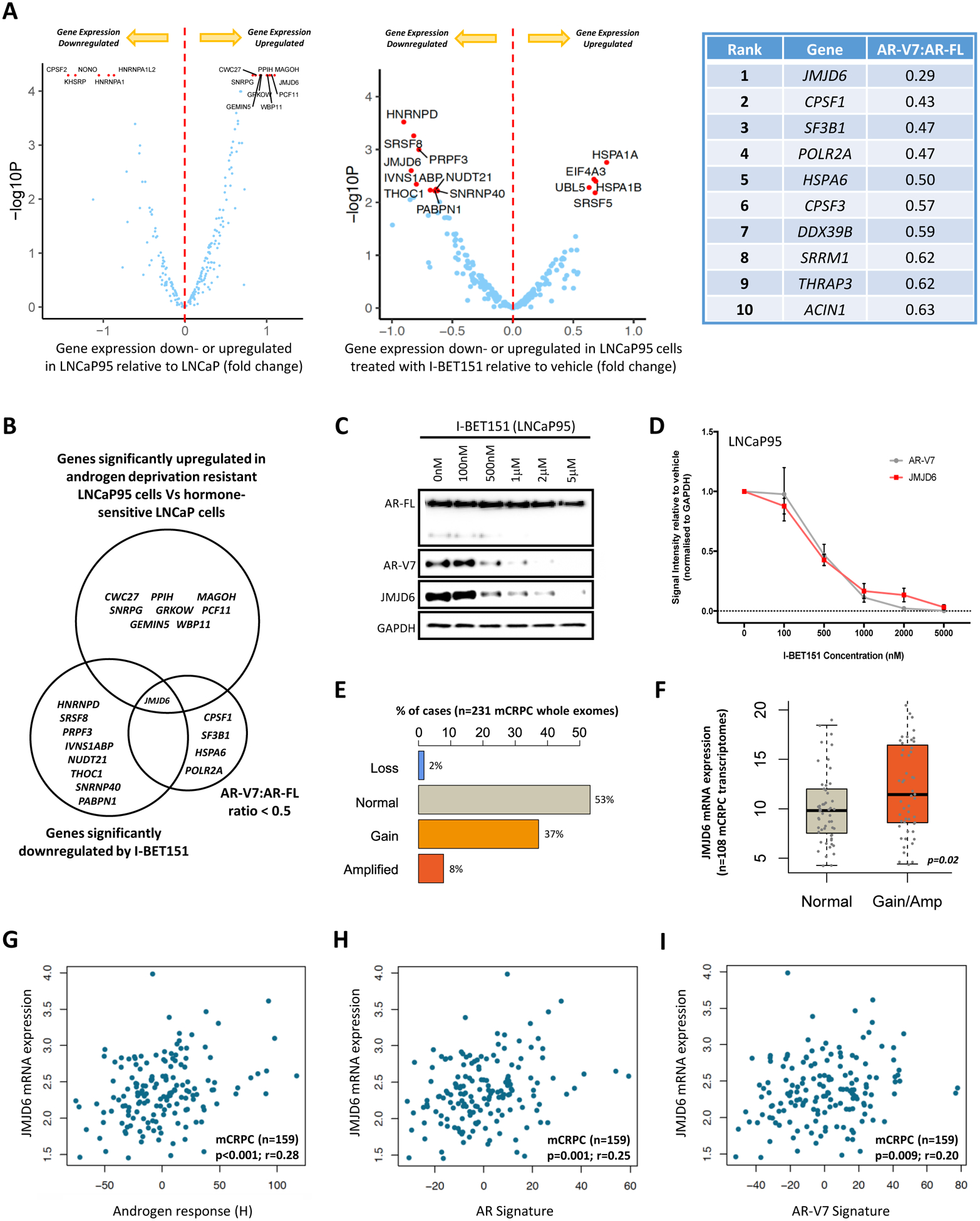
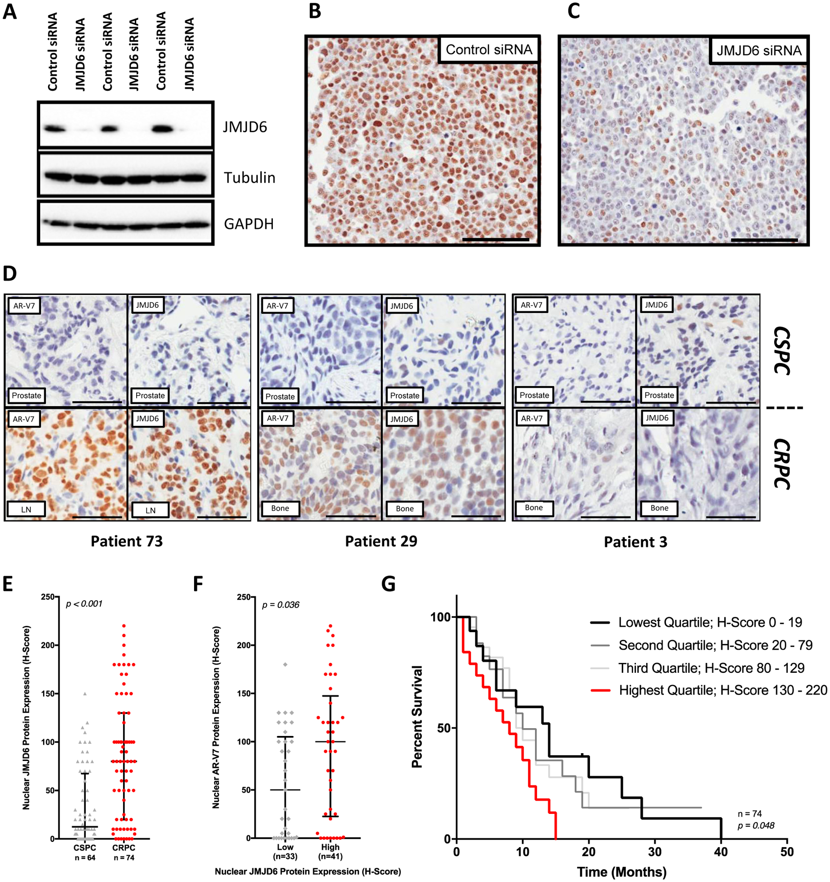
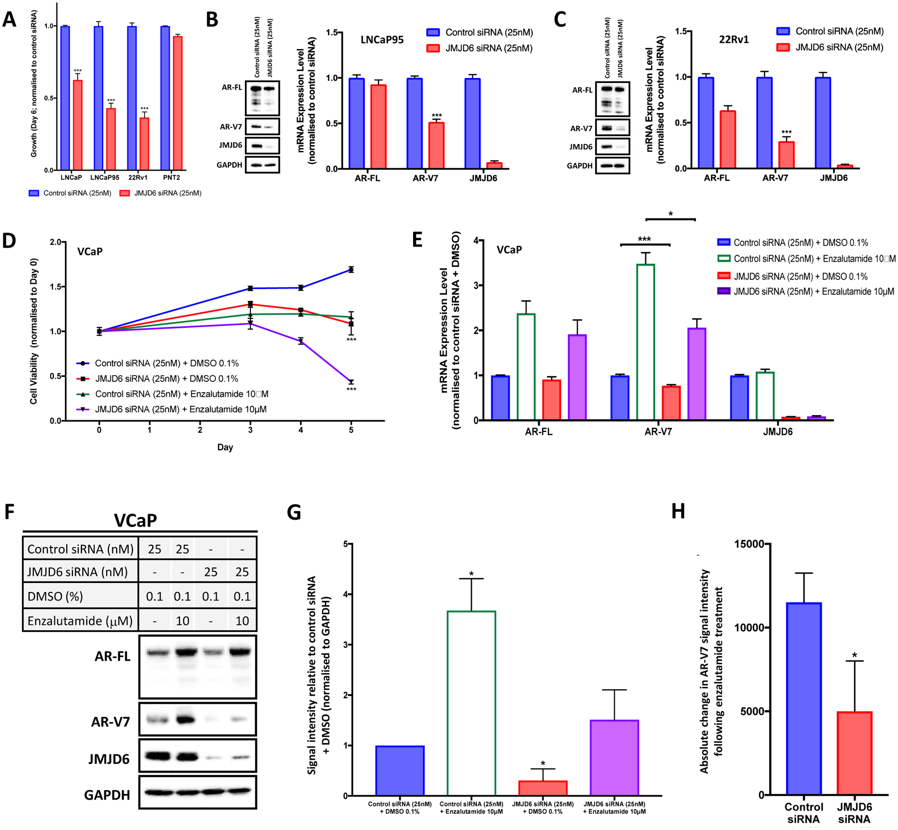
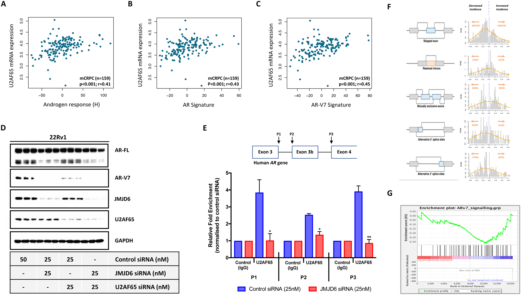
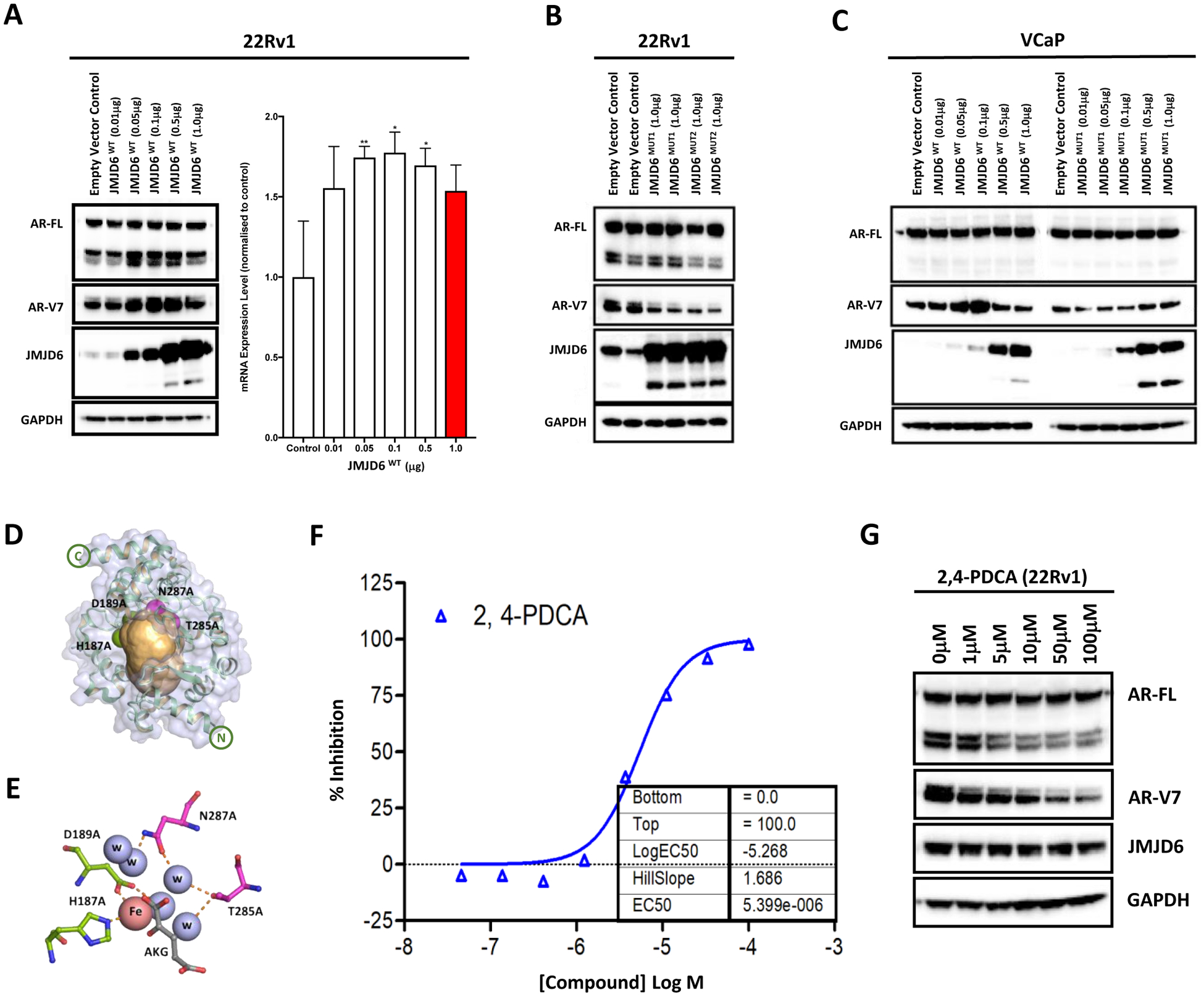
Endocrine resistance (EnR) in advanced prostate cancer is fatal. EnR can be mediated by androgen receptor (AR) splice variants, with AR splice variant 7 (AR-V7) arguably the most clinically important variant. In this study, we determined proteins key to generating AR-V7, validated our findings using clinical samples, and studied splicing regulatory mechanisms in prostate cancer models. Triangulation studies identified JMJD6 as a key regulator of AR-V7, as evidenced by its upregulation with in vitro EnR, its downregulation alongside AR-V7 by bromodomain inhibition, and its identification as a top hit of a targeted siRNA screen of spliceosome-related genes. JMJD6 protein levels increased (P < 0.001) with castration resistance and were associated with higher AR-V7 levels and shorter survival (P = 0.048). JMJD6 knockdown reduced prostate cancer cell growth, AR-V7 levels, and recruitment of U2AF65 to AR pre-mRNA. Mutagenesis studies suggested that JMJD6 activity is key to the generation of AR-V7, with the catalytic machinery residing within a druggable pocket. Taken together, these data highlight the relationship between JMJD6 and AR-V7 in advanced prostate cancer and support further evaluation of JMJD6 as a therapeutic target in this disease. SIGNIFICANCE: This study identifies JMJD6 as being critical for the generation of AR-V7 in prostate cancer, where it may serve as a tractable target for therapeutic intervention.


Conflict of interest statement

Conflicts of interest

AP, JW, AN, WY, IF, RP, AF, RR, DNR, PDM, BG, RC, VG, LB, MC, SM, MBL, SC, NT, BAL, AS and JdB are all employees of The Institute of Cancer Research (ICR), which has a commercial interest in abiraterone. The ICR operates a Rewards to Inventors scheme through which employees of the ICR may receive financial benefit following commercial licensing. BAL is currently or has been a consultant and received fees from to Astex Pharmaceuticals​, GSK and Difiniens AG (member of Astra Zeneca group)​ and received speakers honorarium from Astellas Pharma. BAL is a former employee of Inpharmatica Ltd. SRP is President of ESSA Pharma (Consultant), ProsTech Inc. AS has received travel support from Sanofi and Roche-Genentech, and speakers honorarium from Astellas Pharma. JdB has served on advisory boards and received fees from many companies including Astra Zeneca, Astellas, Bayer, Boehringer Ingelheim, Cellcentric, Daiichi, Genentech/Roche, Genmab, GSK, Janssen, Merck Serono, Merck Sharp & Dohme, Menarini/Silicon Biosystems, Orion, Pfizer, Qiagen, Sanofi Aventis, Sierra Oncology, Taiho, Vertex Pharmaceuticals. He is an employee of The ICR, which have received funding or other support for his research work from AZ, Astellas, Bayer, Cellcentric, Daiichi, Genentech, Genmab, GSK, Janssen, Merck Serono, MSD, Menarini/Silicon Biosystems, Orion, Sanofi Aventis, Sierra Oncology, Taiho, Pfizer, Vertex, and which has a commercial interest in abiraterone, PARP inhibition in DNA repair defective cancers and PI3K/AKT pathway inhibitors (no personal income). JdB was named as an inventor, with no financial interest, for patent 8,822,438. He has been the CI/PI of many industry sponsored clinical trials.

Figures

Figure 1:
Figure 1:. Orthogonal analyses identify the 2OG-dependent dioxygenase JMJD6 as a potential regulator of AR-V7.
(A) Volcano plots illustrating differential mRNA expression of 315 genes relating to the spliceosome (spliceosome related gene set; supplementary table 6), as determined by RNA-seq, between hormone-sensitive LNCaP (no AR-V7 protein) and androgen deprivation resistant LNCaP95 (detectable AR-V7 protein) prostate cancer (PC) cell lines, and LNCaP95 PC cells treated with either a BET inhibitor (I-BET151) or vehicle (DMSO 0.1%). Blue dots represent genes with baseline expression (FPKM) greater than the median expression level of all 315 genes at baseline across both experiments. Top 15 genes most differentially expressed (FPKM) in each experiment (up- or down-regulated) indicated by red dots. Top 10 hits identified in targeted siRNA screen shown in accompanying table; all 315 genes in the spliceosome related gene set were individually inhibited by siRNA in 22Rv1 and LNCaP95 PC cell lines. Changes in AR-V7 protein levels relative to AR-FL were quantified by western blot (WB) densitometry. AR-V7 downregulation averaged across both cell lines with genes ranked in order of the degree of AR-V7 downregulation relative to AR-FL. (B) Venn diagram amalgamating RNA-seq analyses with siRNA screen results. Genes of interest pre-defined as being upregulated in LNCaP95 cells relative to LNCaP cells, downregulated following BET inhibition, and associated with a > 50% reduction in AR-V7 protein expression (WB) relative to AR-FL following siRNA knockdown. JMJD6 was the only gene to meet all three criteria. (C) WB demonstrating that I-BET151 treatment (48 hours) in LNCaP95 PC cells downregulates both AR-V7 and JMJD6 protein expression in a dose-dependent manner. Single representative WB shown from four separate experiments. (D) Densiometric quantification of JMJD6 (red line) and AR-V7 (grey line) protein levels (n=4; densitometry for each biological replicate normalized to GAPDH and vehicle). Demonstrates that protein levels of both JMJD6 and AR-V7 decrease in a dose-dependent manner following BET inhibition with I-BET151. (E) Whole exome analysis (n=231) shows alterations of the JMJD6 gene locus were detected in 47% of mCRPC biopsies (SU2C/PCF cohort), with these being predominately gains (37%; n=86) or amplifications (8%; n=18). (F) Whole exome analysis of mCRPC patients with matched transcriptome data from SU2C/PCF cohort (n=108) shows that JMJD6 copy number gain and amplification (Amp) associated with an increase in JMJD6 mRNA expression in mCRPC biopsies compared to samples without JMJD6 copy number gain/amplification (p=0.02; Wilcoxon test). (G-I) Scatter plots of transcriptome analysis in 159 mCRPC biopsies (SU2C/PCF cohort) showing correlations between JMJD6 mRNA expression and (G) androgen response (Hallmark; H), (H) AR signature (derived from 43 AR regulated transcripts) and (I) AR-V7 signature (derived from 59 genes associated with AR-V7 expression in mCRPC). JMJD6 mRNA expression shown as log FPKM. r-values and p-values are shown and were calculated using Spearman’s correlation.
Figure 2:
Figure 2:. JMJD6 associates with AR-V7 expression and a worse prognosis in mCRPC.
(A) Antibody specificity confirmed by detection of a single band in LNCaP95 whole cell lysates by WB, with downregulation following treatment with pooled JMJD6 siRNA compared to non-targeting control siRNA. (B) Micrograph of LNCaP95 PC cells treated with non-targeting control siRNA demonstrating positive brown nuclear staining for JMJD6. (C) Micrograph of LNCaP95 PC cells treated with pooled JMJD6 siRNA. Demonstrates a marked reduction in JMJD6 protein, with predominately blue, negative staining for JMJD6. (D) Micrographs of IHC analyses for AR-V7 (left) and JMJD6 (right) protein levels in matched, same-patient, diagnostic castration-sensitive (CSPC) (top) and mCRPC (bottom) tissue samples from three different patients (RMH/ICR patient cohort). Scale bars set to 100μm. JMJD6 protein levels in presented tissue samples are similar to AR-V7 levels in mCRPC. (E) Box and whisker plot demonstrating a significant increase (p<0.001) in JMJD6 protein levels (IHC H-Score) in mCRPC biopsies (median H-score [IQR]; CSPC (n=64) 12.5 [0.0–67.5] vs CRPC (n=74) 80 [20.0–130.0]; Wilcoxon rank-sum analysis). (F) Levels of AR-V7 protein significantly higher (p=0.036) in mCRPC tissue samples from patients with high (JMJD6 H-Score ≥ median) mCRPC JMJD6 protein levels (Low 50 [0.0–105.0; n = 33] vs High 100 [22.5–147.5; n = 41]; Mann-Whitney test). (G) Median OS from the time of CRPC tissue biopsy significantly worse in patients with the highest levels of JMJD6 (H-Score > 75th percentile) in their mCRPC tissue sample (n=74, p=0.048; Log-rank test).
Figure 3:
Figure 3:. JMJD6 is important for PC cell growth and regulates AR-V7 expression.
(A) JMJD6 siRNA knockdown (25nM; red bars) significantly reduces the growth (cell number; sulforhodamine B (SRB) assay) of LNCaP, LNCaP95 and 22Rv1 PC cells compared to non-targeting control siRNA (25nM; blue bars), while PNT2 cells (immortalized normal prostatic epithelial cells) were relatively unaffected. Mean cell growth (normalized to control siRNA at same concentration) shown with standard error of the mean; n ≥ 4 data points (at least 2 biological replicates with 2 technical replicates). (B-C) JMJD6 siRNA knockdown downregulates AR-V7 mRNA (qPCR) and protein (WB) levels in LNCaP95 and 22Rv1 PC cell lines. Mean RNA expression (normalized to housekeeping genes (B2M and GAPDH) and control siRNA at equivalent concentration; defined as 1.0) with standard error of the mean from three experiments is shown. (D) Line graph illustrating the impact of JMJD6 siRNA knockdown (25nM) +/− enzalutamide (10μM) on the viability of hormone-sensitive, AR amplified and AR-V7 producing VCaP PC cells compared to controls after five days, as determined using the CellTiter-Glo® Luminescent Cell Viability Assay. JMJD6 siRNA knockdown (red line) significantly reduced VCaP PC cell viability compared to control siRNA (blue line). Combination treatment with enzalutamide (purple line) resulted in a significantly more profound reduction of VCaP cell viability than either JMJD6 siRNA alone (red) or enzalutamide alone (green). n=3; mean cell viability (normalized to control siRNA at same concentration + DMSO 0.1%) shown with standard error of the mean. (E) JMJD6 knockdown downregulated baseline AR-V7 mRNA (qPCR) levels in VCaP cells. JMJD6 knockdown also resulted in a significantly lower increase in AR-V7 mRNA expression in response to AR blockade (enzalutamide 10μM; purple bar) compared to non-targeting control siRNA (green bar). Mean RNA expression (normalized to housekeeping genes (B2M, GAPDH and CDC73), and control siRNA at equivalent concentration + DMSO 0.1%; defined as 1.0) with standard error of the mean from three experiments is shown. (F-G) Single representative WB shown from three separate experiments with corresponding densiometric quantification of AR-V7 protein levels (n=3; densitometry for each biological replicate normalized to GAPDH and vehicle). JMJD6 siRNA knockdown reduces AR-V7 protein levels in VCaP PC cells. Furthermore, while AR-V7 protein levels increase significantly with AR blockade (enzalutamide 10μM), AR-V7 protein levels do not significantly change when JMJD6 is knocked down by siRNA (25nM) at the time of treatment with enzalutamide (10μM). (H) Bar chart showing absolute change in AR-V7 WB signal intensity following AR blockade (enzalutamide 10μM) compared to vehicle (DMSO) with either a non-targeting control siRNA, or a JMJD6 specific siRNA. Demonstrates that the upregulation of AR-V7 in response to enzalutamide was significantly less when JMJD6 was knocked down compared to control siRNA. p values (*, p ≤ 0.05; **, p ≤ 0.01; ***, p ≤ 0.001) were calculated for each condition compared to control (at equivalent concentration) using the mean value of technical replicates with unpaired Student’s t tests.
Figure 4:
Figure 4:. JMJD6 regulates AR-V7 transcription, in part, through recruitment of splicing factor U2AF65 to AR-V7 specific splice sites in in vitro models of CRPC.
(A-C) Scatter plots showing correlations between JMJD6 mRNA expression and (A) androgen response (Hallmark; H), (B) AR signature (derived from 43 AR regulated transcripts) and (C) AR-V7 signature (derived from 59 genes associated with AR-V7 expression in mCRPC) in 159 mCRPC biopsies (SU2C/PCF cohort). U2AF65 mRNA expression shown as log FPKM. r-values and p-values are shown and were calculated using Spearman’s correlation. (D) Single WB in technical triplicate demonstrating reduction in AR-V7 protein levels with both JMJD6 and U2AF65 siRNA in 22Rv1 PC cells. JMJD6 siRNA had minimal impact on U2AF65 protein levels. (E) Schematic diagram of the human AR gene illustrating the regions targeted in RNA immunoprecipitation (RIP) assay with accompanying summary bar chart. Shows a reduction in detectable U2AF65 at the AR-V7 specific splice sites P1 (containing the 5’ splice site for both AR and AR-V7) and P2 (containing the 3’ splice site for AR-V7) in 22Rv1 PC cells treated with JMJD6 siRNA compared to non-targeting control siRNA. Indicates that JMJD6 regulates recruitment of the splicing factor U2AF65 to AR-V7 splice sites. RIP data derived from two independent experiments conducted in triplicate. p values (*, p ≤ 0.05; **, p ≤ 0.01; ***, p ≤ 0.001) were calculated for each condition compared to control (at equivalent concentration) using the mean value of technical replicates with unpaired Student’s t tests. (F) Schematic representation of alternative splicing events alongside corresponding histogram of alternative splicing mean differences between non-targeting control siRNA (blue dotted line; defined as 0.0) and JMJD6 siRNA in LNCaP95 PC cells. Left shift denotes decrease in splicing events. Total number of alternative splicing events (x) occurring in total number of genes (y) shown in orange (x/y). JMJD6 knockdown led to substantial changes in 753 alternative splicing events, with the majority of these occurring less frequently. (G) JMJD6 knockdown in LNCaP95 PC cells associated with a reduction in AR-V7 activity (derived from 59 genes associated with AR-V7 expression in mCRPC); Enrichment Score (ES) = −0.32.
Figure 5:
Figure 5:. Evidence JMJD6-mediated AR-V7 generation is dependent on JMJD6 catalysis, which can be chemically inhibited to downregulate AR-V7 protein levels.
(A) Transfection of a JMJD6 wild-type (JMJD6WT) plasmid at increasing concentrations (all receiving 1μg of plasmid in total, with empty vector control added to make up the difference) into 22Rv1 PC cells led to an increase in AR-V7 protein (WB) and mRNA (qPCR) levels. Mean mRNA levels were normalized to housekeeping genes (B2M and GAPDH), and to studies with an empty vector control plasmid at equivalent concentration; the empty vector control data were defined as 1.0 with standard error of the mean from three experiments shown. p values (*, p ≤ 0.05; **, p ≤ 0.01; ***, p ≤ 0.001) were calculated for each condition compared to control (at equivalent concentration), using the mean value of technical replicates with unpaired Student’s t tests. (B) Conversely, transfection with inactivating mutations of active site residues in the JMJD6 catalytic domain by JMJD6MUT1 (D189A and H187A) and JMJD6MUT2 (N287A and T285A) decreased AR-V7 protein levels (empty vector control, JMJD6MUT1 and JMJD6MUT2 = 1μg of total plasmid). (C) AR-V7 expression was induced by JMJD6WT but not by JMJD6MUT1 in VCaP PC cells, suggesting that JMJD6-mediated AR-V7 expression requires active JMJD6. Singleton WB validating findings presented in (B) in an alternative cell line model. (D-E) Graphic representation of JMJD6 tertiary structure [58]. The inactivating substitutions of active site residues in the JMJD6 catalytic domain by JMJD6MUT1 (D189A and H187A; green spheres) and JMJD6MUT2 (N287A and T285A; magenta spheres) reside within a predicted druggable pocket (shown in orange), identified by the canSAR knowledgebase [33, 34]. (F) Liquid chromatography-mass spectrometry (LC-MS) analysis demonstrating that the 2OG mimic pyridine-2,4-dicarboxylic acid (2,4-PDCA) resulted in a dose-dependent reduction in isolated JMJD6-mediated lysyl-5-hydroxylation of its known target LUC7L; indicating that 2,4-PDCA is an inhibitor of JMJD6 lysyl hydroxylase catalytic activity. (G) WB showing that 2,4-PDCA caused a dose-dependent reduction of AR-V7 protein levels in 22Rv1 PC cells. Single representative WB shown from two separate experiments.

References

    1. Visakorpi T, et al., In vivo amplification of the androgen receptor gene and progression of human prostate cancer. Nat Genet, 1995. 9(4): p. 401–6. - PubMed
    1. Robinson D, et al., Integrative clinical genomics of advanced prostate cancer. Cell, 2015. 161(5): p. 1215–1228. - PMC - PubMed
    1. de Bono JS, et al., Abiraterone and increased survival in metastatic prostate cancer. N Engl J Med, 2011. 364(21): p. 1995–2005. - PMC - PubMed
    1. Scher HI, et al., Increased survival with enzalutamide in prostate cancer after chemotherapy. N Engl J Med, 2012. 367(13): p. 1187–97. - PubMed
    1. Fizazi K, et al., Abiraterone plus Prednisone in Metastatic, Castration-Sensitive Prostate Cancer. N Engl J Med, 2017. 377(4): p. 352–360. - PubMed

Publication types

MeSH terms