. 2021 May 3;153(5):e201912540.
doi: 10.1085/jgp.201912540.

Kir4.1/Kir5.1 channels possess strong intrinsic inward rectification determined by a voltage-dependent K+-flux gating mechanism

Affiliations

Kir4.1/Kir5.1 channels possess strong intrinsic inward rectification determined by a voltage-dependent K+-flux gating mechanism

Leticia G Marmolejo-Murillo et al. J Gen Physiol. .

Abstract

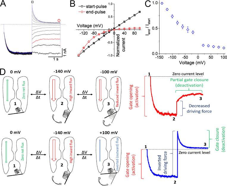
Inwardly rectifying potassium (Kir) channels are broadly expressed in both excitable and nonexcitable tissues, where they contribute to a wide variety of cellular functions. Numerous studies have established that rectification of Kir channels is not an inherent property of the channel protein itself, but rather reflects strong voltage dependence of channel block by intracellular cations, such as polyamines and Mg2+. Here, we identify a previously unknown mechanism of inward rectification in Kir4.1/Kir5.1 channels in the absence of these endogenous blockers. This novel intrinsic rectification originates from the voltage-dependent behavior of Kir4.1/Kir5.1, which is generated by the flux of potassium ions through the channel pore; the inward K+-flux induces the opening of the gate, whereas the outward flux is unable to maintain the gate open. This gating mechanism powered by the K+-flux is convergent with the gating of PIP2 because, at a saturating concentration, PIP2 greatly reduces the inward rectification. Our findings provide evidence of the coexistence of two rectification mechanisms in Kir4.1/Kir5.1 channels: the classical inward rectification induced by blocking cations and an intrinsic voltage-dependent mechanism generated by the K+-flux gating.


Figures

Figure 1.
Figure 1.
Inward rectification of Kir4.1 and Kir4.1/Kir5.1 channels in whole-cell control conditions. (A and C) Macroscopic whole-cell Kir4.1/Kir5.1 (A) and Kir4.1 (C) currents in HEK-293 cells. Currents were evoked by using the voltage-step protocol shown in the inset at the center of the four panels. For clarity, currents at potentials −70, −50, −30, and −10 mV are not shown. The inset of A corresponds to the amplification of the first 50 ms of outward currents from this panel. The dashed line denotes the zero-current level. (B and D) Normalized I-V relationships for Kir4.1/Kir5.1 (B) and Kir4.1 (D) currents measured at the end of the test pulses and normalized to the amplitude of the current recorded at −120 mV. n = 9 (Kir4.1/Kir5.1) and n = 6 (Kir4.1).
Figure 2.
Figure 2.
Heteromeric Kir4.1/Kir5.1 but not homomeric Kir4.1 channels inwardly rectify in inside-out configuration. (A–E) Representative macroscopic Kir4.1/Kir5.1 (A, C, and D) and Kir4.1 (E) currents recorded in the inside-out configuration when applying the triple-pulse voltage protocol depicted in the inset. The bath (intracellular) solution contained 5 mM (A and E), 1 mM (C), or 10 mM (D) EDTA. (B and F) Normalized I-V relationships for Kir4.1/Kir5.1 (B) and Kir4.1 (F) currents measured at the end of the test pulses and normalized to the amplitude of the current recorded at −140 mV. The dashed line denotes the zero-current level. n = 6–8 cells for each channel and EDTA condition.
Figure 3.
Figure 3.
Effect of exogenous SPM on inside-out Kir4.1/Kir5.1 and Kir4.1 currents. (A) Kir4.1/Kir5.1 currents before (control) and after perfusing 1 µM SPM in the bath. Currents were recorded in the inside-out configuration applying the same triple-pulse voltage protocol as for Fig. 2. (B) Normalized I-V relationships for Kir4.1/Kir5.1 in the absence (control) and presence of 1 µM SPM. Currents were measured at the end of the test pulses and normalized to the amplitude of the current recorded at −140 mV in control conditions. (C) Superimposed Kir4.1/Kir5.1 current traces from A illustrating control currents (black traces) and their respective SPM-induced block (red traces) at +40, +60, +80, and +100 mV. (D) Time constants (τslows] and τfastf]) of Kir4.1/Kir5.1 currents as shown in C, in control conditions and in the presence of 1 µM SPM. (E and F) Current recordings (E) and normalized I-V relationships (F) for Kir4.1 in control and after 1 µM SPM, obtained as described for A and B, respectively. The dashed line denotes the zero-current level. Data are presented as mean ± SEM. n = 5 for Kir4.1/Kir5.1 and n = 11 for Kir4.1. *, P = 0.047; +, P = 0.032; x, P = 0.029.
Figure 4.
Figure 4.
Voltage-independent activation and deactivation kinetics of Kir4.1/Kir5.1 channels. (A and C) Inside-out Kir4.1/Kir5.1 current traces elicited with the voltage-step protocols (see insets), which are described in the text and designed for activation (A) or deactivation (C) of currents. (B and D) Time constants of the activation (τ act; B) and deactivation (τ deact; D) of Kir4.1/Kir5.1 currents as a function of the test pulse potential. Time constants in black and red were obtained with single and double exponential functions, respectively. The dashed line denotes the zero-current level. Data are presented as mean ± SEM. n = 8 for activation and n = 6 for deactivation.
Figure 5.
Figure 5.
Exogenous PIP2 largely reduces the inward rectification of Kir4.1/Kir5.1 channels. (A and B) Representative Kir4.1/Kir5.1 current traces elicited by the voltage protocol shown at the bottom of A, in control conditions (A) and in the presence of 10 µM PIP2 (B). (C) Normalized I-V relationships for currents measured at the end of the test pulses, before and after PIP2 perfusion (n = 7). Currents were normalized to that obtained at −140 mV. For comparison, an ohmic behavior is depicted by a dashed line. (D) Superimposed Kir4.1/Kir5.1 current recordings in the presence of 10 µM PIP2 and after adding 10 µM SPM (similar results were obtained in four more cells). Currents were elicited to −140 mV for 2 s previous to a depolarizing pulse (2 s) to +100 mV (holding potential = 0 mV). The horizontal dashed lines denote the zero-current level.
Figure 6.
Figure 6.
Effect of asymmetrical K+ gradient (200 versus 77 mM and 200 versus 19 mM) on Kir4.1/Kir5.1 I-V relationships. (AC, F, and G) Inside-out current traces from Kir4.1/Kir5.1 channels obtained using solutions containing the indicated K+ concentration. Currents were induced by voltage protocols as described in the text. (D and H) Normalized I–V relationships for currents evoked under conditions displayed in A (red circles), B (black line), C (blue triangles), F (maroon squares), and G (green circles). Note: Three additional test pulses are shown in H (empty marron squares) to illustrate the persistence of the inward rectification. Current amplitudes were measured at the end of the test pulses and normalized to the respective maximum value at each condition. Current traces in B are those from Fig. 2, which are only shown for comparison purposes, as well as the respective I-V relationship (continuous line in D). The inset at the right of D refers to the amplification of the area delimited in the box. (E and I) Current ratios for those obtained at the more depolarized and hyperpolarized potentials under the K+ concentration shown in A (red bar), C (blue bar), F (maroon bar), and G (green bar) measuring currents at the end of the test pulses. The dashed line denotes the zero-current level. Data are presented as mean ± SEM. n = 5–6 for all assayed conditions. *, P = 0.048; ***, P = 0.0002.
Figure 7.
Figure 7.
Gating model for Kir4.1/Kir5.1 channel. (A) Representative Kir4.1/Kir5.1 currents as described for Fig. 2. The red and blue traces display the currents when the test pulses were −100 and +100 mV, respectively (which are shown at the right of D to illustrate the channel gating behavior). (B) Normalized I-V relationships measured at the start and at the end of the test pulses and normalized to the amplitude of the current recorded at −140 mV. (C) Ratio Iend/Istart as a function of the test pulse potentials. n = 8 cells. (D) Hypothetical model of the Kir4.1/Kir5.1 gating mechanism operated by the K+-flux. For clarity, two of the four channel subunits are depicted with the ion permeation path between them. The channel activation gate is represented by a hinge. According to the model, inward currents elicited by hyperpolarizing pulses induce the opening of the gate, whereas depolarizing pulses deactivate (to varying degrees) the channel. The partial deactivation of the channels observed at voltages negative to EK could be either because the gate partially closes or due to a decrease in the channel open probability. Please see a detailed explanation of the hypothetical model in the Discussion section.

Comment in

References

    1. Aleksandrov, A., Velimirovic B., and Clapham D.E.. 1996. Inward rectification of the IRK1 K+ channel reconstituted in lipid bilayers. Biophys. J. 70:2680–2687. 10.1016/S0006-3495(96)79837-5 - DOI - PMC - PubMed
    1. Bellot-Saez, A., Kékesi O., Morley J.W., and Buskila Y.. 2017. Astrocytic modulation of neuronal excitability through K+ spatial buffering. Neurosci. Biobehav. Rev. 77:87–97. 10.1016/j.neubiorev.2017.03.002 - DOI - PubMed
    1. Chen, T.Y., and Miller C.. 1996. Nonequilibrium gating and voltage dependence of the ClC-0 Cl- channel. J. Gen. Physiol. 108:237–250. 10.1085/jgp.108.4.237 - DOI - PMC - PubMed
    1. De Jesús-Pérez, J.J., Castro-Chong A., Shieh R.C., Hernández-Carballo C.Y., De Santiago-Castillo J.A., and Arreola J.. 2016. Gating the glutamate gate of CLC-2 chloride channel by pore occupancy. J. Gen. Physiol. 147:25–37. 10.1085/jgp.201511424 - DOI - PMC - PubMed
    1. Fakler, B., Brändle U., Bond C., Glowatzki E., König C., Adelman J.P., Zenner H.P., and Ruppersberg J.P.. 1994. A structural determinant of differential sensitivity of cloned inward rectifier K+ channels to intracellular spermine. FEBS Lett. 356:199–203. 10.1016/0014-5793(94)01258-X - DOI - PubMed

Publication types