Review
. 2021 Sep:225:107848.
doi: 10.1016/j.pharmthera.2021.107848. Epub 2021 Apr 3.

Targeting autophagy in ischemic stroke: From molecular mechanisms to clinical therapeutics

Affiliations
Review

Targeting autophagy in ischemic stroke: From molecular mechanisms to clinical therapeutics

Amir Ajoolabady et al. Pharmacol Ther. 2021 Sep.

Abstract

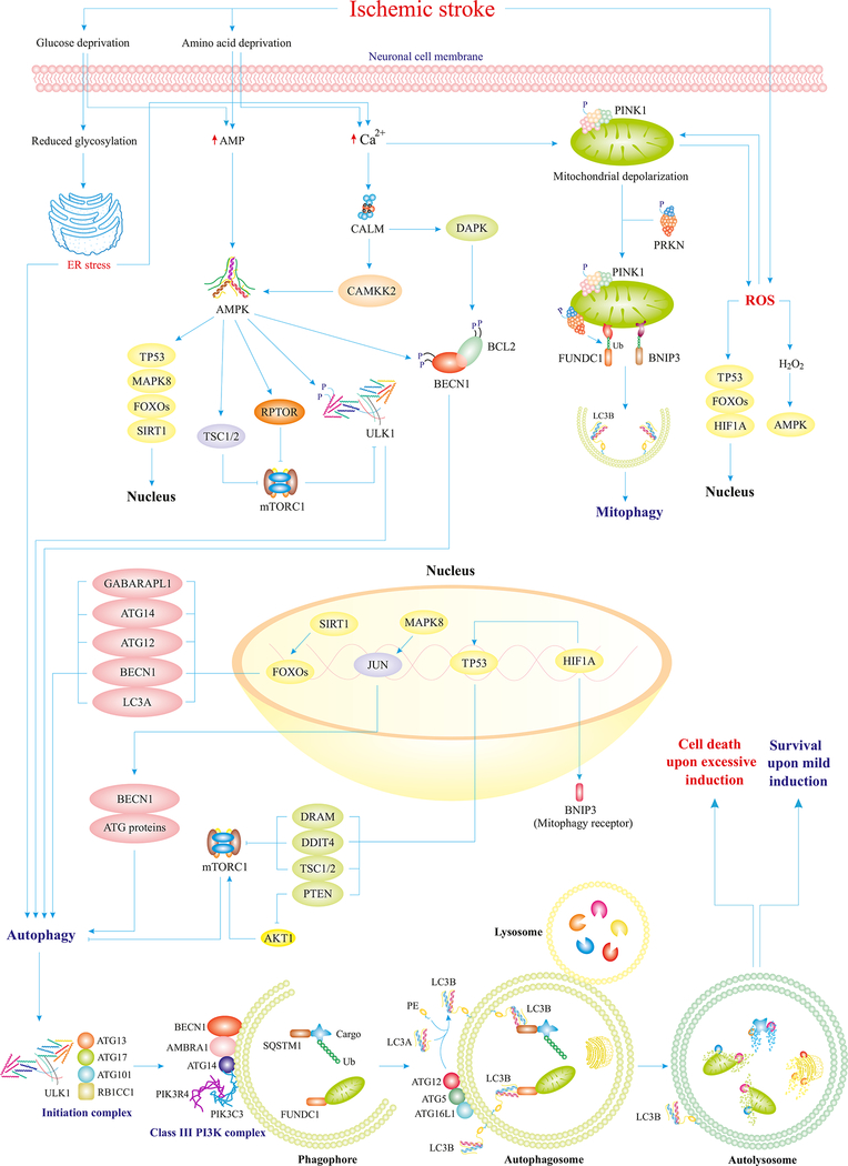
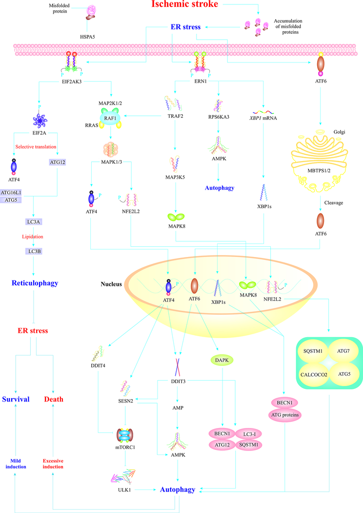
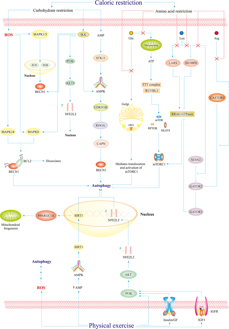
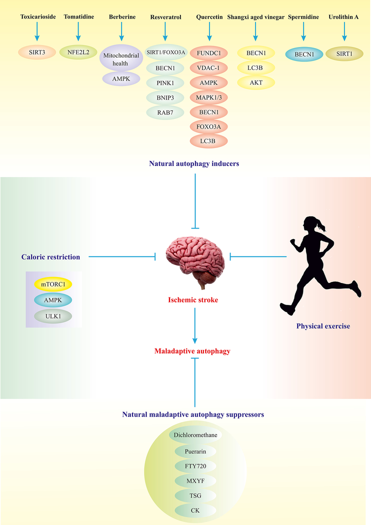
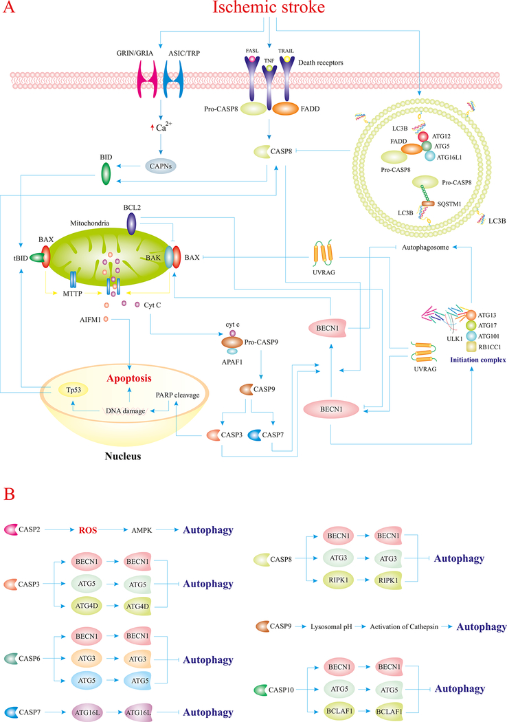
Stroke constitutes the second leading cause of death and a major cause of disability worldwide. Stroke is normally classified as either ischemic or hemorrhagic stroke (HS) although 87% of cases belong to ischemic nature. Approximately 700,000 individuals suffer an ischemic stroke (IS) in the US each year. Recent evidence has denoted a rather pivotal role for defective macroautophagy/autophagy in the pathogenesis of IS. Cellular response to stroke includes autophagy as an adaptive mechanism that alleviates cellular stresses by removing long-lived or damaged organelles, protein aggregates, and surplus cellular components via the autophagosome-lysosomal degradation process. In this context, autophagy functions as an essential cellular process to maintain cellular homeostasis and organismal survival. However, unchecked or excessive induction of autophagy has been perceived to be detrimental and its contribution to neuronal cell death remains largely unknown. In this review, we will summarize the role of autophagy in IS, and discuss potential strategies, particularly, employment of natural compounds for IS treatment through manipulation of autophagy.

Keywords: Adaptive autophagy; Cell death; Cerebral I/R injury; Ischemic stroke; Maladaptive autophagy.


Figures

Fig. 1.
Fig. 1.
IS perturbs autophagy homeostasis through distinct mechanisms. IS provokes deprivation of oxygen and nutrient as a result of interrupted blood flow. Loss of glucose and amino acids overtly promotes accumulation of AMP, Ca2+, and ROS in the cytosol of neurons. Overproduction of AMP turns on the master regulator of autophagy, AMPK, which facilitates upregulation of ATG proteins, inhibition of mTORC1, and activation of ULK1. Elevated levels of Ca2+ also activate AMPK and depolarize mitochondrial membrane, which would in turn evoke mitophagy and ROS production. In response to ROS generation, AMPK and transcriptional factors are activated to further promote ATG proteins and mitophagy receptors. Ultimately, activated ATG proteins pave the way for the initiation of autophagy to foster the clearance of long-lived or damaged organelles, and superfluous proteins through the autophagosomal-lysosomal fusion. ULK1-ATG13-ATG101-RB1CC1/FIP200 form the autophagy initiation complex, BECN1-NRBF2-ATG14-PIK3R4/VPS15-PIK3C3/VPS34 form the class III phosphatidylinositol 3-kinase complex, which aids the initiation complex in the formation of phagophores, and the ATG12–ATG5-ATG16L1 complex mediates lipidation of LC3A and formation of autophagosomes around damaged organelles or certain cargos. Excessive induction of autophagy results in cell death (ACD/autosis), whereas normal/mild induction improves neuronal survival in IS (Ajoolabady, Aghanejad, et al., 2020; Ghislat & Knecht, 2015; Mrakovcic & Fröhlich, 2018; J. Ren, et al., 2020; Simon, Friis, Tait, & Ryan, 2017). Abbreviations: CAMKK2 (calcium/calmodulin dependent protein kinase kinase 2), DAPK (death associated protein kinase), Ub (ubiquitin), FUNDC1 (FUN14 domain containing 1), RPTOR/raptor (regulatory associated protein of mTOR complex 1), JUN/C-Jun (Jun proto-oncogene, AP-1 transcription factor subunit), GABARAPL1 (GABA type A receptor associated protein like 1), DRAM (DNA damage regulated autophagy modulator), DDIT4 (DNA damage inducible transcript 4), RB1CC1/FIP200 (RB1 inducible coiled-coil 1), NRBF2 (nuclear receptor binding factor 2), PIK3R4/VPS15 (phosphoinositide-3-kinase regulatory subunit 4), UVRAG (UV radiation resistance associated gene), PE (phosphatidylethanolamine).
Fig. 2.
Fig. 2.
IS induces ER stress, which triggers autophagy through three major ER stress sensors. IS-induced ER stress mediates EIF2AK3 dimerization and activation. EIF2AK3 phosphorylates/activates EIF2A/eIF2α, which induces reticulophagy (selective autophagy of the ER) via selective translation of ATG12 leading to alleviation of ER stress. EIF2AK3 primarily induces adaptive autophagy via activation of ATF4 and NFE2L2/Nrf-2 proteins, which translocate to the nucleus and induce autophagy via distinct machineries. AMPK activation, upregulation of BECN1 and ATG genes, and mTORC1 inhibition are the main mechanisms of ATF4-mediated autophagy. NFE2L2/Nrf-2 also induces autophagy through upregulation of autophagy-associated proteins. Upon ER stress insult, ERN1 also undergoes autophosphorylation and homodimerization. Activated ERN1 induces activation of MAPK8 which translocates to the nucleus and induces autophagy via the upregulation of BECN1 and ATG genes. The endoribonuclease domain of ERN1 cleaves XBP1 mRNA, which produces XBP1s that translocates to the nucleus and induces autophagy through upregulation of BECN1 and ATG genes. The third branch of UPR, ATF6, also translocates to the Golgi and gets cleaved by MBTPS1/S1P and MBTPS2/S2P, which produces activated ATF6 which translocates to the nucleus, triggers adaptive responses, and upregulates DAPK, to evoke autophagy. Autophagy initially acts as an adaptive response element that relieves ER stress. However, severe ER stress induces maladaptive autophagy, leading to neuronal cell death (Amir Ajoolabady, et al., 2021; Fusakio, et al., 2016; Jurkin, et al., 2014; Papaioannou, et al., 2018; Radanovic, et al., 2020; Urra, et al., 2018; D. Wang, et al., 2018; Yucel, et al., 2019) Abbreviations: HSPA5/Bip (heat shock protein family A (Hsp70) member 5), MAP2K½(mitogen-activated protein kinase kinase ½), RRAS (RAS related), RAF1 (Raf-1 proto-oncogen, serine/threonine kinase), EIF2A/eIF2α (eukaryotic translation initiation factor 2A), TRAF2 (TNF receptor associated factor 2), MAP3K5 (mitogen-activated protein kinase kinase kinase 5), RPS6KA3 (ribosomal protein S6 kinase A3), XBP1 (X-box binding protein 1), MBTPS½ (membrane bound transcription factor peptidase, site ½), MAPK3/ERK1 (mitogen-activated protein kinase 3), XBP1s (spliced X-box binding protein 1), ATF4 (activating transcription factor 4), DDIT3/CHOP (DNA damage inducible transcript 3), SESN2 (sestrin 2), AMP (adenosine monophosphate).
Fig. 3.
Fig. 3.
Caloric restriction and physical exercise maintain adaptive autophagy. Carbohydrate restriction induces mild generation of ROS in the cytosol, which activates the MAPK1/ERK2-MAPK3/ERK1 pathways leading to the upregulation of BECN1. Besides, IKK activation mediates activation of potential autophagy regulators and elements such as AMPK, BECN1, and MAPK8. The PI3K-AKT1-NFE2L2/Nrf-2 pathway also leads to nuclear translocation of NFE2L2/Nrf-2 and autophagy activation. Both carbohydrate and amino acid restriction culminate in accelerated AMP levels in the cytosol and subsequent AMPK activation. Reduced Gln uptake blocks Golgi-mediated mTORC1 activation and ATP-mediated formation of mTORC1 components. Leu restriction mediates SH3BP4 activation and LARS/LRS and GATOR2 inhibition all of which culminates in autophagy induction via suppression of the RRAG GTPases-mTORC1 cascade. Similarly, Arg restriction also places a stop codon on GATOR2, which results in GATOR1 activation and subsequent blockade of RRAG GTPases and mTORC1, and, thus, autophagy activation. Physical exercise also mediates autophagy activation via reduced cytosolic AMP level, activation of the PI3K-AKT1-NFE2L2/Nrf-2 pathway, and mild ROS generation. Abbreviations: IKK (inhibitor of nuclear factor-kappa B kinase subunit beta), Gln (Glutamine), Leu (leucine), Arg (arginine), SH3BP4 (SH3 domain-binding protein 4), LARS/LRS (leucyl-tRNA synthetase), STK11/LKB1 (serine/threonine kinase 11), FOS/C-Fos (Fos proto-oncogene, transcription factor subunit), CDKN1B/p27/Kip1 (cyclin dependent kinase inhibitor 1B), TTT (Tel2-Tti1-Tti2) complex, RUVBL1 (RuvB like AAA ATPase 1), CASTOR1 (cytosolic arginine sensor for mTORC1 subunit 1), RRAG (Ras related GTPl binding), MLST8 (mTOR associated protein, LST8 homolog), ARF1 (ADP ribosylation factor 1), MAPK14/p38 (mitogen-activated protein kinase 14), CAPN (calpains protein family), GATOR2 (GTPase activating proteins toward Rags subcomplex 2), GATOR1 (Gap Activity Toward Rags 1), NFE2L2/Nrf-2 (nuclear factor, erythroid 2 like 2), PPARGC1A/PGC1-α (PPARG coactivator 1 alpha), GF (growth factor), IGFR (insulin like growth factor 1 receptor).
Fig. 4.
Fig. 4.
Strategies for manipulation of autophagy in IS management. Depicted natural compounds have the potential to induce adaptive autophagy or block maladaptive autophagy via manipulation of autophagy elements. Certain natural autophagy inducers have been reported to mildly turn on several autophagy regulators, leading to adaptive autophagy and alleviation of IS. Physical exercise and caloric restriction are the most suitable, conventional and applicable measures to maintain adaptive autophagy and alleviate IS.(Ajoolabady, Aslkhodapasandhokmabad, Aghanejad, Zhang, & Ren, 2020) Abbreviations: SIRT3 (sirtuin 3), RAB7 (RAB7, member RAS oncogene family), VDAC1 (voltage dependent anion channel 1).
Fig. 5.
Fig. 5.
Crosstalk between autophagy and apoptosis in IS Abbreviations: FADD (Fas associated via death domain), CASP8 (caspase 8), BID (BH3 interacting domain death agonist), MTTP (microsomal triglyceride transfer protein), CASP9 (caspase 9), APAF1 (apoptotic peptidase activating factor), GRIN (glutamate ionotropic receptor NMDA type subunits), GRIA (glutamate ionotropic receptor AMPA type subunits), CASP7 (caspase 7), CASP2 (caspase 2), CASP10 (caspase 10).

Similar articles

Cited by

References

    1. Aidar FJ, de Oliveira RJ, Silva AJ, de Matos DG, Mazini Filho ML, Hickner RC, & Machado Reis V (2012). The influence of resistance exercise training on the levels of anxiety in ischemic stroke. Stroke research and treatment, 2012. - PMC - PubMed
    1. Ajoolabady A, Aghanejad A, Bi Y, Zhang Y, Aslkhodapasand HH, Abhari A, & Ren J (2020). Enzyme-based autophagy in anti-neoplastic management: From molecular mechanisms to clinical therapeutics. Biochimica et Biophysica Acta (BBA)-Reviews on Cancer, 188366. - PubMed
    1. Ajoolabady A, Aslkhodapasandhokmabad H, Aghanejad A, Zhang Y, & Ren J (2020). Mitophagy Receptors and Mediators: Therapeutic Targets in the Management of Cardiovascular Ageing. Ageing Research Reviews, 101129. - PubMed
    1. Ajoolabady A, Aslkhodapasandhokmabad H, Henninger N, Demillard LJ, Nikanfar M, Nourazarian A, & Ren J (2021). Targeting Autophagy in Neurodegenerative Diseases: from Molecular Mechanisms to Clinical Therapeutics. Clin Exp Pharmacol Physiol. - PMC - PubMed
    1. Ajoolabady A, Wang S, Kroemer G, Klionsky DJ, Uversky VN, Sowers JR, Aslkhodapasandhokmabad H, Bi Y, Ge J, & Ren J (2021). ER stress in Cardiometabolic Diseases: from Molecular Mechanisms to Therapeutics. Endocrine Reviews. - PubMed

Publication types

MeSH terms