

Strange eyes, stranger brains: exceptional diversity of optic lobe organization in midwater crustaceans
- PMID: 33823669
- PMCID: PMC8059609
- DOI: 10.1098/rspb.2021.0216
Strange eyes, stranger brains: exceptional diversity of optic lobe organization in midwater crustaceans
Abstract
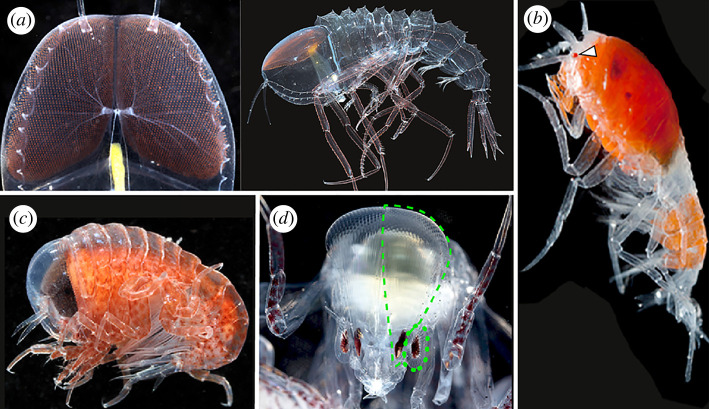
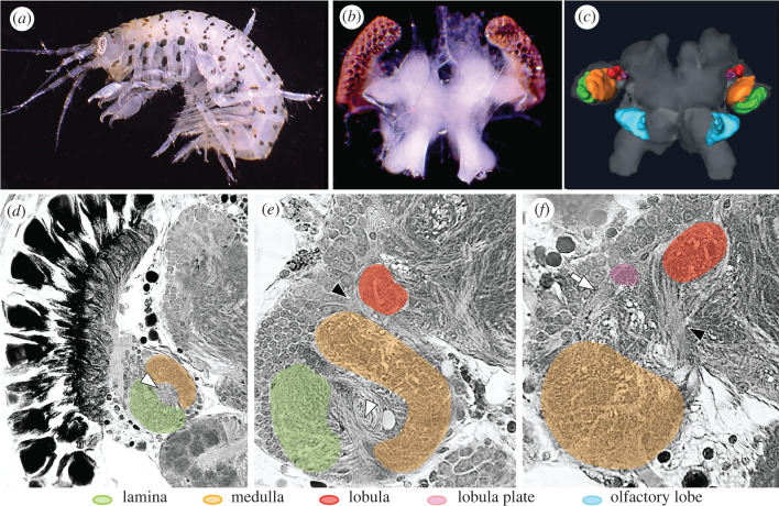
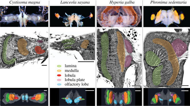
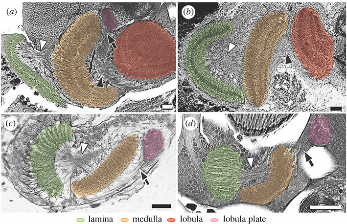
Nervous systems across Animalia not only share a common blueprint at the biophysical and molecular level, but even between diverse groups of animals the structure and neuronal organization of several brain regions are strikingly conserved. Despite variation in the morphology and complexity of eyes across malacostracan crustaceans, many studies have shown that the organization of malacostracan optic lobes is highly conserved. Here, we report results of divergent evolution to this 'neural ground pattern' discovered in hyperiid amphipods, a relatively small group of holopelagic malacostracan crustaceans that possess an unusually wide diversity of compound eyes. We show that the structure and organization of hyperiid optic lobes has not only diverged from the malacostracan ground pattern, but is also highly variable between closely related genera. Our findings demonstrate a variety of trade-offs between sensory systems of hyperiids and even within the visual system alone, thus providing evidence that selection has modified individual components of the central nervous system to generate distinct combinations of visual centres in the hyperiid optic lobes. Our results provide new insights into the patterns of brain evolution among animals that live under extreme conditions.
Keywords: brain evolution; compound eyes; hyperiid amphipods; neuroanatomy; optic lobes.
Figures
Similar articles
-
Convergent evolution of optic lobe neuropil in Pancrustacea.Arthropod Struct Dev. 2021 Mar;61:101040. doi: 10.1016/j.asd.2021.101040. Epub 2021 Mar 9. Arthropod Struct Dev. 2021. PMID: 33706077 Review.
-
Phylogenetic analysis of lineage relationships among hyperiid amphipods as revealed by examination of the mitochondrial gene, cytochrome oxidase I (COI).Integr Comp Biol. 2007 Dec;47(6):815-30. doi: 10.1093/icb/icm093. Epub 2007 Oct 30. Integr Comp Biol. 2007. PMID: 21669761
-
Brain organization and the origin of insects: an assessment.Proc Biol Sci. 2009 Jun 7;276(1664):1929-37. doi: 10.1098/rspb.2008.1471. Epub 2009 Feb 25. Proc Biol Sci. 2009. PMID: 19324805 Free PMC article. Review.
-
Complex brain and optic lobes in an early Cambrian arthropod.Nature. 2012 Oct 11;490(7419):258-61. doi: 10.1038/nature11495. Nature. 2012. PMID: 23060195
-
Molecular phylogenetic evidence for the reorganization of the Hyperiid amphipods, a diverse group of pelagic crustaceans.Mol Phylogenet Evol. 2013 Apr;67(1):28-37. doi: 10.1016/j.ympev.2012.12.021. Epub 2013 Jan 11. Mol Phylogenet Evol. 2013. PMID: 23319084
Cited by
-
Bioimaging marine crustacean brain: quantitative comparison of micro-CT preparations in an Alpheid snapping shrimp.Front Neurosci. 2024 Nov 26;18:1428825. doi: 10.3389/fnins.2024.1428825. eCollection 2024. Front Neurosci. 2024. PMID: 39659887 Free PMC article.
-
Multiple axes of visual system diversity in Ithomiini, an ecologically diverse tribe of mimetic butterflies.J Exp Biol. 2023 Dec 15;226(24):jeb246423. doi: 10.1242/jeb.246423. Epub 2023 Dec 8. J Exp Biol. 2023. PMID: 37921078 Free PMC article.
-
Matched function of the neuropil processing optic flow in flies and crabs: the lobula plate mediates optomotor responses in Neohelice granulata.Proc Biol Sci. 2022 Aug 31;289(1981):20220812. doi: 10.1098/rspb.2022.0812. Epub 2022 Aug 17. Proc Biol Sci. 2022. PMID: 35975436 Free PMC article.
References
-
- Robison BH. 2004. Deep pelagic biology. J. Exp. Mar. Biol. Ecol. 300, 253-272. (10.1016/j.jembe.2004.01.012) - DOI
-
- Zeidler W. 2012. A review of the hyperiidean amphipod families Mimonectidae and Proscinidae (Crustacea: Amphipoda: Hyperiidea: Scinoidea). Zootaxa 3533, 1-74. (10.11646/zootaxa.3533.1.1) - DOI
-
- Land MF. 2000. Eyes with mirror optics. J. Opt. A: Pure Appl. Opt. 2, R44-R50. (10.1088/1464-4258/2/6/204) - DOI
-
- Land MF, Nilsson D-E. 2012. Animal eyes, 2nd ed. New York, NY: Oxford University Press.
Publication types
MeSH terms
Associated data
LinkOut - more resources
Full Text Sources
Other Literature Sources
