

Adipose-derived stem cells combined with platelet-rich plasma enhance wound healing in a rat model of full-thickness skin defects
- PMID: 33823915
- PMCID: PMC8022317
- DOI: 10.1186/s13287-021-02257-1
Adipose-derived stem cells combined with platelet-rich plasma enhance wound healing in a rat model of full-thickness skin defects
Abstract
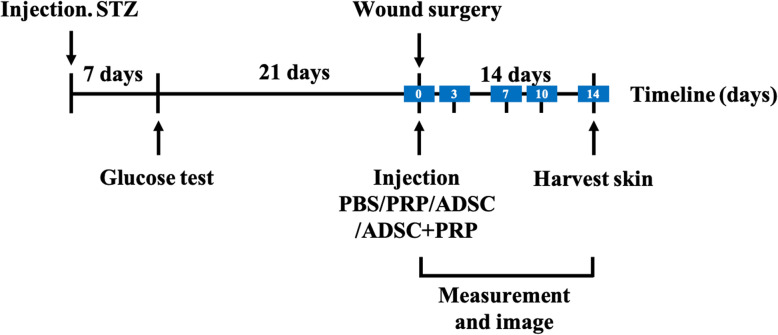
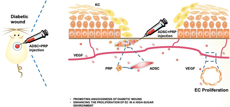
Background: Wound healing is impaired in patients with diabetes due to the multifactorial etiology of the disease, which limits the therapeutic efficacy of various approaches. This study hypothesizes that the combination of adipose-derived stem cells (ADSCs) and platelet-rich plasma (PRP) might achieve optimally efficient diabetic wound healing.
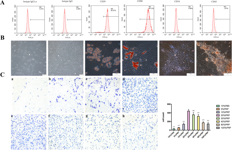
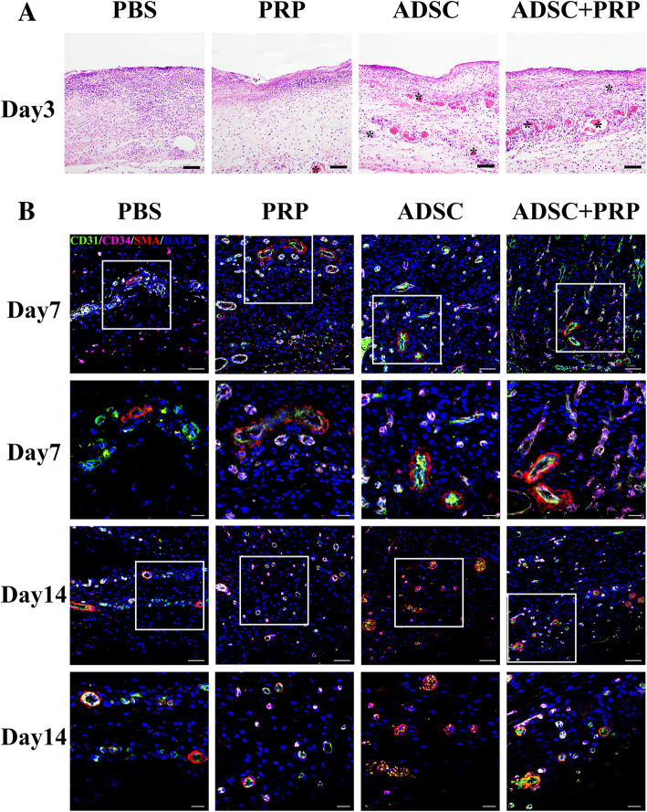
Methods: ADSCs were isolated from the adipose tissues of Sprague-Dawley (SD) rats. PRP was prepared by using a two-step centrifugation technique. A diabetic wound model was established on the backs of SD rats to evaluate the effect of ADSCs incorporated into PRP. Hematoxylin and eosin staining, immunofluorescence, and immunohistochemistry were performed to observe the changes in neovascularization. ELISA and Western blot were utilized to detect the angiogenesis-related protein expression levels. The proliferation of endothelial cells was assessed by the MTS assay.
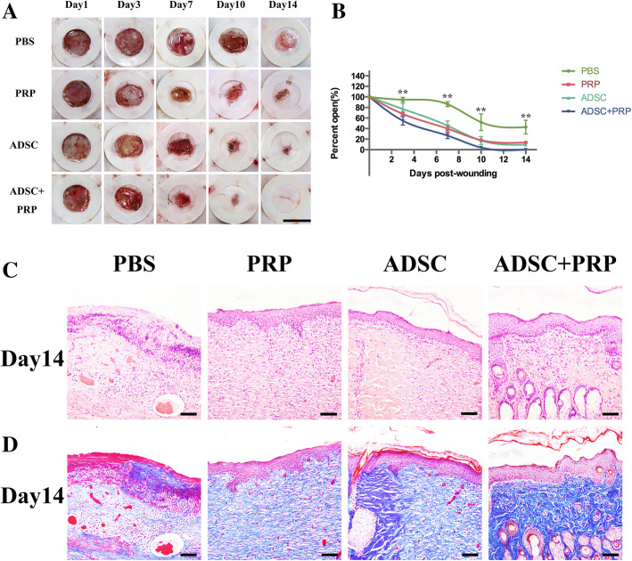
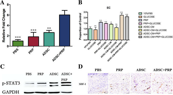
Results: ADSCs incorporated into PRP induced a higher wound closure rate than ADSCs, PRP, and negative control. The expression levels of VEGF, p-STAT3, and SDF-1 in the ADSC+PRP group were higher than those in the other groups. Moreover, the proliferation of endothelial cells was strongly stimulated by treatment with the combination of ADSC-conditioned medium (ADSC-CM) and PRP.
Conclusions: PRP enhanced diabetic wound healing induced by ADSCs, and its promoting effect involved neovascularization.
Keywords: Adipose-derived stem cell; Diabetic wound healing; Neovascularization; Platelet-rich plasma.
Conflict of interest statement
The authors declare that they have no competing interests.
Figures
Similar articles
-
[Effects of human adipose-derived mesenchymal stem cells and platelet-rich plasma on healing of wounds with full-thickness skin defects in mice].Zhonghua Shao Shang Za Zhi. 2018 Dec 20;34(12):887-894. doi: 10.3760/cma.j.issn.1009-2587.2018.12.013. Zhonghua Shao Shang Za Zhi. 2018. PMID: 30585053 Chinese.
-
Adipose mesenchymal stem cells combined with platelet-rich plasma accelerate diabetic wound healing by modulating the Notch pathway.Stem Cell Res Ther. 2021 Jul 13;12(1):392. doi: 10.1186/s13287-021-02454-y. Stem Cell Res Ther. 2021. PMID: 34256844 Free PMC article.
-
[Effects of hypoxia-pretreated rat adipose-derived mesenchymal stem cells conditioned medium on wound healing of rats with full-thickness defects].Zhonghua Shao Shang Za Zhi. 2020 Sep 20;36(9):803-812. doi: 10.3760/cma.j.cn501120-20200508-00258. Zhonghua Shao Shang Za Zhi. 2020. PMID: 32972065 Chinese.
-
Fat grafting and platelet-rich plasma in wound healing: a review of histology from animal studies.Adipocyte. 2021 Dec;10(1):80-90. doi: 10.1080/21623945.2021.1876374. Adipocyte. 2021. PMID: 33525977 Free PMC article. Review.
-
The use of fat grafting and platelet-rich plasma for wound healing: A review of the current evidence.Int Wound J. 2019 Feb;16(1):275-285. doi: 10.1111/iwj.13029. Epub 2018 Nov 20. Int Wound J. 2019. PMID: 30460739 Free PMC article. Review.
Cited by
-
Ameliorative Effect of Combined Placenta-Derived Mesenchymal Stem Cells plus Platelet-rich Plasma on Polycystic Ovarian Model in Rats: A Biochemical and Histological Study.Reprod Sci. 2025 Mar;32(3):907-918. doi: 10.1007/s43032-025-01791-0. Epub 2025 Jan 24. Reprod Sci. 2025. PMID: 39856459
-
Overexpression of miR-192 in fibroblasts accelerates wound healing in diabetic rats: research article.Eur J Med Res. 2025 Apr 4;30(1):239. doi: 10.1186/s40001-025-02449-y. Eur J Med Res. 2025. PMID: 40186269 Free PMC article.
-
Low-glucose culture environment can enhance the wound healing capability of diabetic adipose-derived stem cells.Stem Cell Res Ther. 2023 Sep 4;14(1):236. doi: 10.1186/s13287-023-03478-2. Stem Cell Res Ther. 2023. PMID: 37667384 Free PMC article.
-
Optimization of Novel Human Acellular Dermal Dressing Sterilization for Routine Use in Clinical Practice.Int J Mol Sci. 2021 Aug 6;22(16):8467. doi: 10.3390/ijms22168467. Int J Mol Sci. 2021. PMID: 34445173 Free PMC article.
-
Wound-Healing Potential of Myristica fragrans Essential Oil: A Multi-Targeted Approach Involving Inflammation, Oxidative Stress, and Apoptosis Regulation.Pharmaceuticals (Basel). 2025 Jun 12;18(6):880. doi: 10.3390/ph18060880. Pharmaceuticals (Basel). 2025. PMID: 40573275 Free PMC article.
References
Publication types
MeSH terms
LinkOut - more resources
Full Text Sources
Other Literature Sources
Research Materials
Miscellaneous
