. 2021 Apr 6;14(677):eabd2319.
doi: 10.1126/scisignal.abd2319.

Input-specific regulation of glutamatergic synaptic transmission in the medial prefrontal cortex by mGlu2/mGlu4 receptor heterodimers

Affiliations

Input-specific regulation of glutamatergic synaptic transmission in the medial prefrontal cortex by mGlu2/mGlu4 receptor heterodimers

Zixiu Xiang et al. Sci Signal. .

Abstract

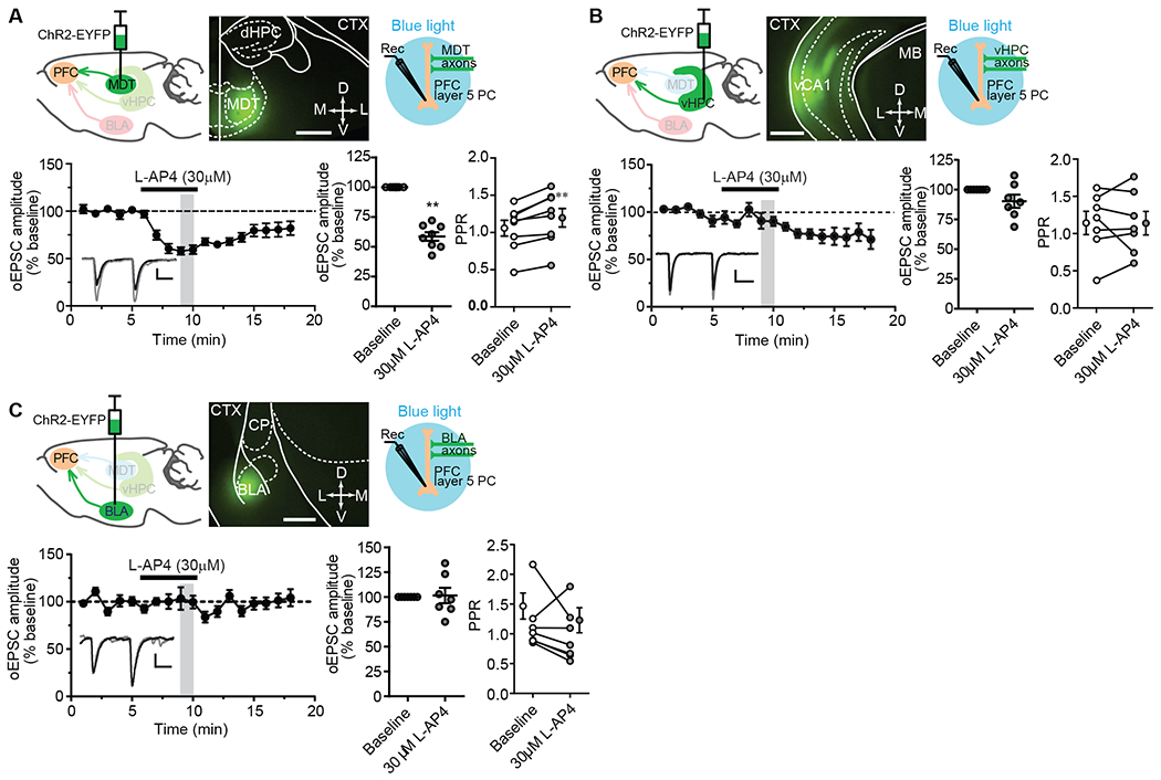
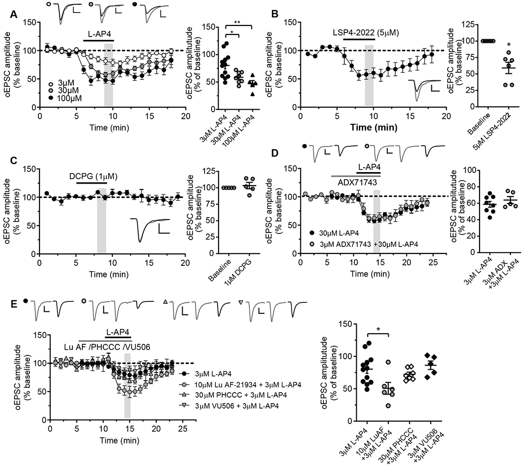
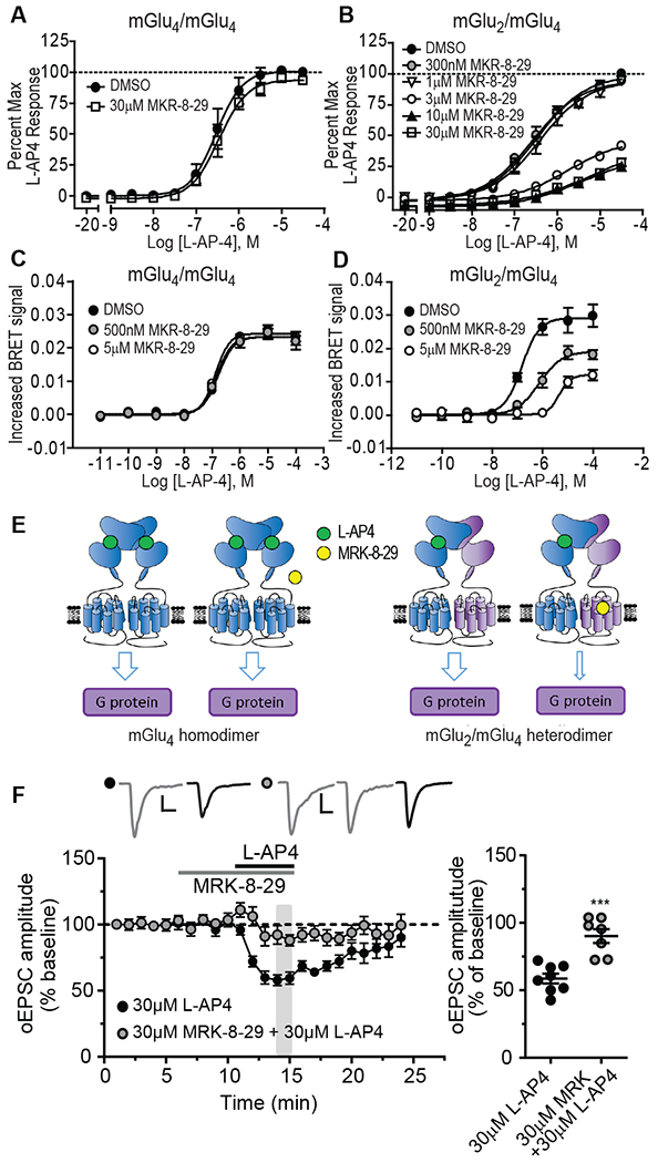
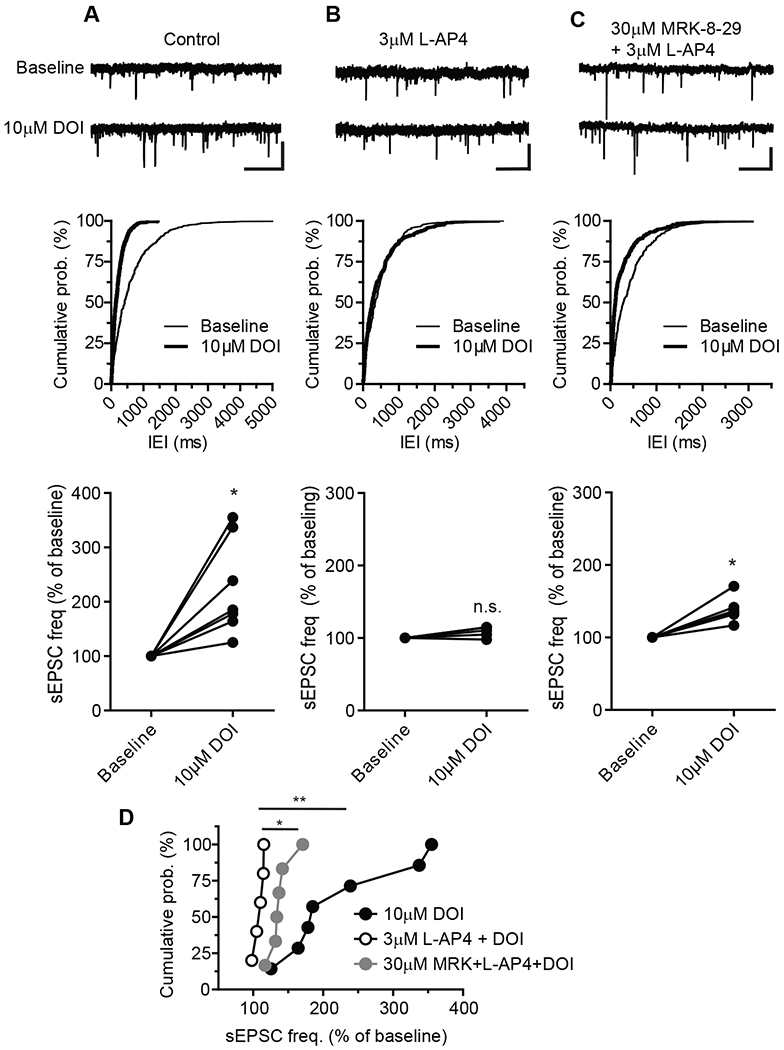
Metabotropic glutamate receptors (mGluRs) are G protein-coupled receptors that regulate various aspects of central nervous system processing in normal physiology and in disease. They are thought to function as disulfide-linked homodimers, but studies have suggested that mGluRs can form functional heterodimers in cell lines. Using selective allosteric ligands, ex vivo brain slice electrophysiology, and optogenetic approaches, we found that two mGluR subtypes-mGluR2 and mGluR4 (or mGlu2 and mGlu4)-exist as functional heterodimers that regulate excitatory transmission in a synapse-specific manner within the rodent medial prefrontal cortex (mPFC). Activation of mGlu2/mGlu4 heterodimers inhibited glutamatergic signaling at thalamo-mPFC synapses but not at hippocampus-mPFC or amygdala-mPFC synapses. These findings raise the possibility that selectively targeting these heterodimers could be a therapeutic strategy for some neurologic and neuropsychiatric disorders involving specific brain circuits.


Conflict of interest statement

Competing interests: D.E.O. is currently an employee of AbbVie, Inc. The other authors declare that they have no competing interests.

Figures

Fig. 1.
Fig. 1.. Effects of L-AP4 on optogenetically evoked glutamatergic transmission at thalamo-, hippocampo-, and amygdala-mPFC synapses.
(A to C) Schematic diagram showing the injection of AAV5-CaMKIIa-hChR2(E123T/T159C)-EYFP into the mediodorsal thalamic nucleus (MDT; A), ventral hippocampus (vHPC; B), or basolateral amygdala (BLA; C), each with a representative image of EYFP-tagged ChR2 expression in the injection site 3 to 5 weeks after viral injection (calibration bar, 200μm), and a second schematic (right) depicting the acquisition of ex vivo whole-cell recordings from mPFC layer V pyramidal cells in response to light stimulation of ChR2:EYFP expressed exons. Bottom panels show the effect of L-AP4 on both oEPSCs over time and paired-pulse ratios (PPR) at each synapse type. Data are mean ± SEM from N = 8 neurons from total of 7 mice (A), and 7 neurons from 5 (B) or 4 (C) mice. Synapse-specific oEPSCs versus baselines were compared by Wilcoxon matched-pairs tests: **P <0.01 (A), P > 0.1 (B), and P > 0.9 (C). PPRs were also compared by Wilcoxon matched-pairs tests: **P < 0.01 (A), P > 0.9 (B), and P > 0.2 (C). Abbreviations: dHPC = dorsal hippocampus, CTX = cortex, vCA1= ventral CA1, MB = midbrain, CP = caudal putamen. Calibration bars for oEPSC traces: 100pA/20ms (A), 50pA/20ms (B), and 20pA/20ms (C). The gray-shaded vertical bar in the time course indicates the time points used to be averaged for statistical comparisons to baseline. For reference but not marked in the figure is the statistical comparison of oEPSCs and PPR at 14-15 min after L-AP4 application at hippocampo-mPFC synapses (B): P < 0.05 and P > 0.9, respectively, by Wilcoxon matched-pairs test.
Fig. 2.
Fig. 2.. Effects of L-AP4 and mGlu4 PAMs with different pharmacologic profiles on glutamatergic transmission at thalamo-mPFC synapse.
(A) Time courses (left) and dot plot (right) summarizing the effects of L-AP4 at different concentration on oEPSCs at thalamo-mPFC synapses (N = 12 neurons from 10 mice for 3 μM L-AP4, 8 neurons from 7 mice for 30 μM L-AP4 [data are from Fig.1A], and 5 neurons from 3 mice for 100 μM L-AP4. *P < 0.05 and **P < 0.01 by one-way ANOVA, [F(2,22)=7.30, P = 0.0037] with Bonferroni’s post-test. (B) The effect of LSP4-2022 on oEPSCs at thalamo-mPFC synapses. N = 6 neurons from 5 mice; *P < 0.05 by Wilcoxon matched-pairs test. (C) The effect of mGlu8 agonist DCPG on oEPSCs. N = 5 neurons from 3 mice; P > 0.4 by Wilcoxon matched-pairs test. (D) The effect of mGlu7 NAM ADX71743 with 30 μM L-AP4 on oEPSCs (N = 5 neurons from 3 mice) compared to the effect of 30 μM L-AP4 alone (same data as in Fig. 1A). P > 0.4 by Mann-Whitney test. (E and F) The effect of mGlu4 PAM Lu AF21934, PHCCC or VU0418506 with 3 μM L-AP4 on oEPSCs (N = 6 neurons from 4 mice, 7 per 5, and 5 per 4, respectively) compared to the effect of 3 μM L-AP4 on oEPSCs (same data as in panel A). *P < 0.05 by one-way ANOVA [F(3,26) = 4.14, P = 0.0159] with Dunnett’s post-test. Calibration bars for oEPSC traces: 100pA/10ms, 100pA/10ms, 40pA/10ms (A); 100pA/10ms (B, C); 200pA/10ms (D); 100pA/10ms (E).
Fig. 3.
Fig. 3.. Effect of mGlu2 NAM MRK-8-29 on L-AP4 induced inhibition of thalamo-mPFC synaptic transmission.
(A and B) The effects of MRK-8-29 on L-AP4 CRC in HEK cells expressing mGlu4 homodimers (A) or mGlu2/4 heterodimers (B) by GIRK assay. (C and D) The effects of MRK-8-29 on L-AP4 CRC in HEK cells expressing mGlu4 homodimers (C) or mGlu2/4 heterodimers (D) by CODA-RET assay. (E) The schematic diagrams illustrate the effect of MRK-8-29 on L-AP4 induced response at mGlu4 homodimers (left) or mGlu2/4 heterodimers (right). (F) The effect of 30 μM MRK-8-29 with L-AP4 on oEPSCs (N = 7 neurons from 5 mice) compared to the effect of 30 μM L-AP4 alone (same data as in Fig. 1A). ***P < 0.001 by Mann-Whitney test. Calibration bars for oEPSC traces: 100pA/10ms.
Fig. 4.
Fig. 4.. Effects of L-AP4 and MRK-8-29 on 5-HT2 agonist DOI induced increase in sEPSCs in mPFC layer V pyramidal cells.
(A to C) Sample traces (top), cumulative probability plots of inter-event interval (IEI) of sEPSCs (middle) from three representative cells and summary plot of normalized group data (bottom) before and during application of 10μM DOI (A), before and during application of 10μM DOI in the presence of 3 μM L-AP4, and before and during application of 10μM DOI in the presence of 30 μM MRK-8-29 and 3 μM L-AP4. Calibration bars for sEPSC traces: 20pA/1s (A and B), and 30pA/1s (C). Normalized sEPSC frequency after application of 10μM DOI verse baseline under different conditions were compared using Wilcoxon matched-pairs tests: *P < 0.05, N = 7 neurons from 5 mice (A, bottom); P > 0.1, N = 5 per 4 (B, bottom); *P < 0.05, N = 6 per 4 (C, bottom). (D) Cumulative probability plots summarizing DOI-induced sEPSC frequency change in control, in the presence of L-AP4 and in the combination of L-AP4 and MRK-8-29. * P < 0.05 and ** P < 0.01 by one-way ANOVA [F(2,15)=7.113, P = 0.0067] with Bonferroni’s post-test.

References

    1. Nicoletti F, Bockaert J, Collingridge GL, Conn PJ, Ferraguti F, Schoepp DD, Wroblewski JT, Pin JP, Metabotropic glutamate receptors: from the workbench to the bedside. Neuropharmacology 60, 1017–1041 (2011); published online Epub6/2011 (S0028-3908(10)00292-3 [pii];10.1016/j.neuropharm.2010.10.022 [doi]). - DOI - PMC - PubMed
    1. Niswender CM, Conn PJ, Metabotropic glutamate receptors: physiology, pharmacology, and disease. Annual review of pharmacology and toxicology 50, 295–322 (2010); published online Epub2010 (10.1146/annurev.pharmtox.011008.145533). - DOI - PMC - PubMed
    1. Nickols HH, Conn PJ, Development of allosteric modulators of GPCRs for treatment of CNS disorders. Neurobiol.Dis 61, 55–71 (2014); published online Epub1/2014 (S0969-9961(13)00259-3 [pii];10.1016/j.nbd.2013.09.013 [doi]). - DOI - PMC - PubMed
    1. Kunishima N, Shimada Y, Tsuji Y, Sato T, Yamamoto M, Kumasaka T, Nakanishi S, Jingami H, Morikawa K, Structural basis of glutamate recognition by a dimeric metabotropic glutamate receptor. Nature 407, 971–977 (2000); published online Epub10/26/2000 (10.1038/35039564 [doi]). - DOI - PubMed
    1. Romano C, Yang WL, O’Malley KL, Metabotropic glutamate receptor 5 is a disulfide-linked dimer. J.Biol.Chem 271, 28612–28616 (1996); published online Epub11/8/1996 ( - PubMed

Publication types

LinkOut - more resources