Review
. 2021 May;21(5):319-329.
doi: 10.1038/s41577-021-00536-9. Epub 2021 Apr 6.

Endothelial dysfunction and immunothrombosis as key pathogenic mechanisms in COVID-19

Affiliations
Review

Endothelial dysfunction and immunothrombosis as key pathogenic mechanisms in COVID-19

Aldo Bonaventura et al. Nat Rev Immunol. 2021 May.

Abstract

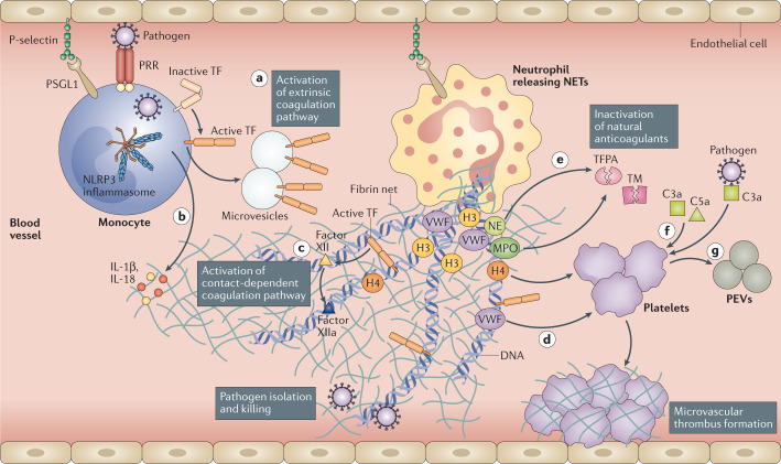
Coronavirus disease 2019 (COVID-19) is a clinical syndrome caused by infection with severe acute respiratory syndrome coronavirus 2 (SARS-CoV-2). Patients with severe disease show hyperactivation of the immune system, which can affect multiple organs besides the lungs. Here, we propose that SARS-CoV-2 infection induces a process known as immunothrombosis, in which activated neutrophils and monocytes interact with platelets and the coagulation cascade, leading to intravascular clot formation in small and larger vessels. Microthrombotic complications may contribute to acute respiratory distress syndrome (ARDS) and other organ dysfunctions. Therapeutic strategies aimed at reducing immunothrombosis may therefore be useful. Several antithrombotic and immunomodulating drugs have been proposed as candidates to treat patients with SARS-CoV-2 infection. The growing understanding of SARS-CoV-2 infection pathogenesis and how it contributes to critical illness and its complications may help to improve risk stratification and develop targeted therapies to reduce the acute and long-term consequences of this disease.


Conflict of interest statement

A.B. and A.V. received a travel grant from Kiniksa Pharmaceuticals Ltd and honoraria from Effetti s.r.l. (Milan, Italy). L.D. has received consultation honoraria from Abbvie, Amgen, Biogen, Bristol-Myers Squibb, Celltrion, GlaxoSmithKline, Novartis, Pfizer, Roche, Sanofi-Genzyme and SOBI. K.M. is an inventor on patent US9642822 and patent applications WO20180271953A1 and US2019167680A1, and is a consultant for PEEL Therapeutics, Inc. B.W.V.T. has served as a consultant for Novartis and Serpin Pharma, and has received research support from Novartis, Kiniksa Pharmaceuticals Ltd, Serpin Pharma and SOBI. A.A. has served as a consultant for Applied Clinical Intel, AstraZeneca, Cromos Pharma, Janssen, Kiniksa Pharmaceuticals Ltd, Merck, Olatec and Serpin Pharma, and has received research support from Janssen, Olatec and Serpin Pharma. All other authors declare no competing interests.

Figures

Fig. 1
Fig. 1. Immunothrombosis is important in promoting immune defence.
Following recognition of a pathogen through pattern recognition receptors (PRRs), monocytes and monocyte-derived microvesicles present activated tissue factor (TF) on their surfaces and release it at sites of pathogen localization, thus activating the extrinsic pathway of coagulation (path a). Pathogens also stimulate the NLRP3 inflammasome in monocytes and/or macrophages, leading to the release of pro-inflammatory cytokines, such as interleukin-1β (IL-1β) and IL-18 (path b). Neutrophils are recruited and contribute to this process through the release of neutrophil extracellular traps (NETs), which directly activate factor XII and, thus, the contact-dependent pathway of coagulation (path c). NETs also bind von Willebrand factor (VWF) and help to recruit platelets (path d). Histones, in particular H3 and H4, trigger activation of platelets. In addition, neutrophil elastase (NE) and myeloperoxidase (MPO) in NETs cleave and inactivate natural anticoagulants (tissue factor pathway inhibitor (TFPA) and thrombomodulin (TM)) (path e). Finally, NETs can externalize and bind TF, promoting activation of the extrinsic pathway of coagulation. Platelets support the immunothrombotic process by activating the contact-dependent pathway of coagulation through the release of polyphosphates and, along with endothelial cells, may promote fibrin generation. Platelets can also be activated by C3a and C5a (path f). Activated platelets release large amounts of pro-inflammatory cytokines in platelet extracellular vesicles (PEVs) (path g). Through this mechanism, pathogens such as severe acute respiratory syndrome coronavirus 2 (SARS-CoV-2) become trapped within the fibrin-based NETs and killed. The immunothrombotic process allows pathogen killing to be restricted to the intravascular compartment, thus limiting injury to organs. Although it is clear that immunothrombosis participates in SARS-CoV-2 pathogenesis, the exact mechanisms are still under investigation. These may include the following: direct injury of endothelial cells by the virus and consequent activation of the coagulation cascade; infiltration of neutrophils that lead to NET formation; induction of hypoxaemia causing upregulation of TF expression by hypoxia-inducible transcription factors and formation of clots; activation of complement that promotes coagulation and recruits and activates platelets, monocytes and neutrophils, thus triggering TF expression; and an abnormal increase in the levels of pro-inflammatory cytokines causing direct cell damage.
Fig. 2
Fig. 2. Seminal pathology features of patients with COVID-19.
A | Hyaline membrane formation is a typical finding in patients with alveolar damage irrespective of being ventilated or not (blue arrows in panels a and b). Organizing fibrosis (blue arrowhead) and fibrin thrombi in small blood vessels (black arrows) with oedema, along with extensive haemorrhage (haematoxylin and eosin staining) and hyaline membranes (blue arrows) (panel c). The bronchial respiratory mucosa is almost entirely intact and no squamous metaplasia is evident (panel d), different to that observed in patients with severe acute respiratory syndrome coronavirus 2 (SARS-CoV-2). B | Two thrombi, one in a small pulmonary artery (blue arrow) and one in a pulmonary venule (blue arrowhead), in the haematoxylin and eosin staining (panel a) and in the CD61 immunostaining for platelets within thrombi (panel b). Megakaryocytes within small vessels and alveolar capillaries (panel c, blue arrow). CD61 immunostaining of a fibrin- and platelet-rich thrombus in a small vessel (panel d), with a megakaryocyte stained below (blue arrow). Small, perivascular aggregates of lymphocytes (panel e). COVID-19, coronavirus disease 2019. Images reprinted from The Lancet, 8, Fox, S. E. et al., Pulmonary and cardiac pathology in African American patients with COVID-19: an autopsy series from New Orleans, 681–686, Copyright (2020), with permission from Elsevier (ref.).

Comment in

References

    1. Pascarella G, et al. COVID-19 diagnosis and management: a comprehensive review. J. Intern. Med. 2020;288:192–206. - PMC - PubMed
    1. Bonaventura A, et al. Targeting GM-CSF in COVID-19 pneumonia: rationale and strategies. Front. Immunol. 2020;11:1625. - PMC - PubMed
    1. McFadyen JD, Stevens H, Peter K. The emerging threat of (micro)thrombosis in COVID-19 and its therapeutic implications. Circ. Res. 2020;127:571–587. - PMC - PubMed
    1. Ranucci M, et al. The procoagulant pattern of patients with COVID-19 acute respiratory distress syndrome. J. Thromb. Haemost. 2020;18:1747–1751. - PMC - PubMed
    1. Gupta A, et al. Extrapulmonary manifestations of COVID-19. Nat. Med. 2020;26:1017–1032. - PMC - PubMed

Publication types

MeSH terms

Substances

LinkOut - more resources