Value of 18F-FDG Hybrid PET/MR in Differentiated Thyroid Cancer Patients with Negative 131I Whole-Body Scan and Elevated Thyroglobulin Levels
- PMID: 33824601
- PMCID: PMC8018385
- DOI: 10.2147/CMAR.S293005
Value of 18F-FDG Hybrid PET/MR in Differentiated Thyroid Cancer Patients with Negative 131I Whole-Body Scan and Elevated Thyroglobulin Levels
Abstract
Purpose: To evaluate the diagnostic performance of 18F-FDG PET/MR in detecting recurrent or metastatic disease in patients with differentiated thyroid cancer (DTC) who have increased thyroglobulin (Tg) levels but a negative 131I whole-body scan (WBS). The relationship between 18F-FDG PET/MR and serum Tg levels was explored. We also evaluated the therapeutic impact of PET/MR on patient clinical management.
Patients and methods: Twenty-nine DTC patients with a negative 131I-WBS of the last post-therapeutic and increased Tg levels under thyroid-stimulating hormone suppression treatment who underwent 18F-FDG PET/MR examination were retrospectively analyzed.
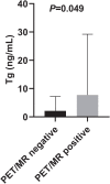
Results: Of those 29 patients, 18F-FDG PET/MR findings were true positive, true negative, false positive, and false negative in 18, 7, 2, and 2 patients, respectively. The overall sensitivity, specificity, and accuracy were 90.0%, 77.8%, and 86.2%, respectively. We noticed significant differences in serum Tg levels between the PET/MR-positive and PET/MR-negative patient groups (P=0.049). Receiver operating characteristic curve analysis showed that a Tg level of 2.4 ng/mL was the optimal cut-off value for predicting PET/MR results. The sensitivity, specificity, and accuracy of PET/MR were higher in patients with Tg levels greater than 2.4 ng/mL than in patients with lower levels. By detecting recurrent or metastatic disease, 18F-FDG PET/MR altered the clinical management in 7 patients (24.1%) of the overall population.
Conclusion: 18F-FDG PET/MR has high diagnostic accuracy for detecting recurrent or metastatic diseases in DTC patients and is useful for clinical management.
Keywords: 18F-FDG; PET/MR; differentiated thyroid cancer; thyroglobulin.
© 2021 Li et al.
Conflict of interest statement
The authors report no conflicts of interest in this work.
Figures


References
-
- Haugen BR, Alexander EK, Bible KC, et al. 2015 American thyroid association management guidelines for adult patients with thyroid nodules and differentiated thyroid cancer: the American thyroid association guidelines task force on thyroid nodules and differentiated thyroid cancer. Thyroid. 2016;26(1):1–133. doi: 10.1089/thy.2015.0020 - DOI - PMC - PubMed
-
- Fatourechi V, Hay ID. Treating the patient with differentiated thyroid cancer with thyroglobulin-positive iodine-131 diagnostic scan-negative metastases: including comments on the role of serum thyroglobulin monitoring in tumor surveillance. Semin Nucl Med. 2000;30(2):107–114. doi: 10.1053/nm.2000.4600 - DOI - PubMed
LinkOut - more resources
Full Text Sources
Other Literature Sources
Miscellaneous

