. 2021 Mar 20:2021:6699054.
doi: 10.1155/2021/6699054. eCollection 2021.

Alleviation of Inflammation and Oxidative Stress in Pressure Overload-Induced Cardiac Remodeling and Heart Failure via IL-6/STAT3 Inhibition by Raloxifene

Affiliations

Alleviation of Inflammation and Oxidative Stress in Pressure Overload-Induced Cardiac Remodeling and Heart Failure via IL-6/STAT3 Inhibition by Raloxifene

Shengqi Huo et al. Oxid Med Cell Longev. .

Retraction in

Abstract

Background: Inflammation and oxidative stress are involved in the initiation and progress of heart failure (HF). However, the role of the IL6/STAT3 pathway in the pressure overload-induced HF remains controversial.

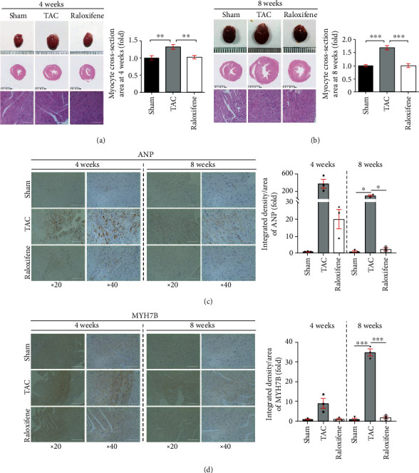
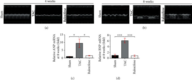
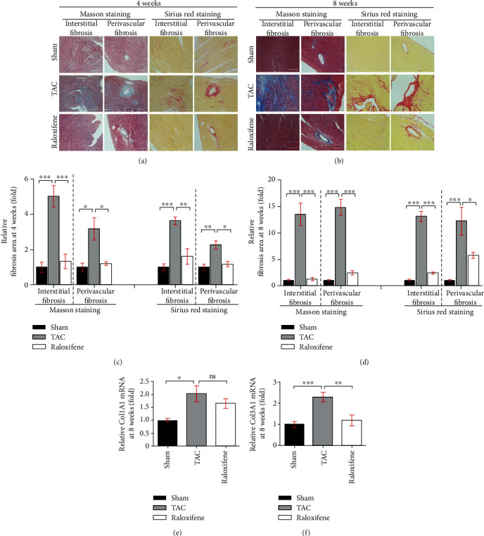
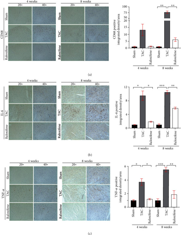
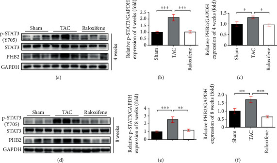
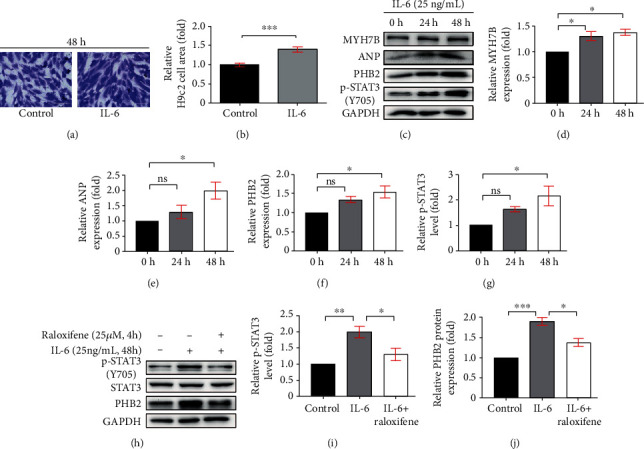
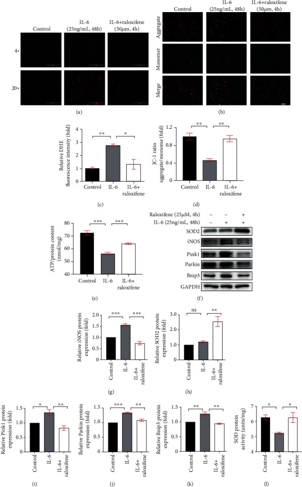
Methods and results: Transverse aortic constriction (TAC) was used to induce pressure overload-HF in C57BL/6J mice. 18 mice were randomized into three groups (Sham, TAC, and TAC+raloxifene, n = 6, respectively). Echocardiographic and histological results showed that cardiac hypertrophy, fibrosis, and left ventricular dysfunction were manifested in mice after TAC treatment of eight weeks, with aggravation of macrophage infiltration and interleukin-6 (IL-6) and tumor necrosis factor-alpha (TNF-α) expression in the myocardium. TAC (four and eight weeks) elevated the phosphorylation of signal transducer and activator of transcription 3 (p-STAT3) and prohibitin2 (PHB2) protein expression. Importantly, IL-6/gp130/STAT3 inhibition by raloxifene alleviated TAC-induced myocardial inflammation, cardiac remodeling, and dysfunction. In vitro, we demonstrated cellular hypertrophy with STAT3 activation and oxidative stress exacerbation could be elicited by IL-6 (25 ng/mL, 48 h) in H9c2 myoblasts. Sustained IL-6 stimulation increased intracellular reactive oxygen species, repressed mitochondrial membrane potential (MMP), decreased intracellular content of ATP, and led to decreased SOD activity, an increase in iNOS protein expression, and increased protein expression of Pink1, Parkin, and Bnip3 involving in mitophagy, all of which were reversed by raloxifene.

Conclusion: Inflammation and IL-6/STAT3 signaling were activated in TAC-induced HF in mice, while sustained IL-6 incubation elicited oxidative stress and mitophagy-related protein increase in H9c2 myoblasts, all of which were inhibited by raloxifene. These indicated IL-6/STAT3 signaling might be involved in the pathogenesis of myocardial hypertrophy and HF.


Conflict of interest statement

The authors declare that they have no conflict of interest.

Figures

Figure 1
Figure 1
Raloxifene mitigated murine cardiac hypertrophy induced by TAC. (a, b) Gross morphology of murine heart, transverse sections, and typical regional enlarged images showed cardiac hypertrophy induced by TAC through HE staining. The respective histogram showed the cross-section area of cardiomyocytes enlarged and raloxifene alleviated the hypertrophic response. (c, d) The increased ANP (c) and MYH7B (d) expression of myocardial tissues after TAC of 4 or 8 weeks detected by immunohistochemistry was mitigated by raloxifene. The minimal interval of scale in the three rows of (a) and (b) is 1 mm, 0.5 mm, and 100 μm, respectively, and 100 μm of (c) and (d). p < 0.05; ∗∗p < 0.01; ∗∗∗p < 0.001.
Figure 2
Figure 2
Raloxifene reversed the cardiac dysfunction of pressure overload mice. (a, b) M-mode echocardiography images of mice measured at 4 (a) or 8 (b) weeks after TAC. (c, d) The increased mRNA expression of ANP (c) and BNP (d) in the murine hearts at 8 weeks were both reduced by raloxifene treatment. p < 0.05; ∗∗∗p < 0.001.
Figure 3
Figure 3
Raloxifene alleviated cardiac fibrosis induced by pressure overload. (a, b) The interstitial and perivascular fibrosis of murine hearts increased by Masson staining (left two columns) and Sirius red staining (right two columns) after TAC of 4 (a) or 8 (b) weeks and was alleviated by raloxifene (scale bar, 200 μm). (c, d) The respective histogram exhibited the quantitation for the cardiac fibrosis area of murine hearts at 4 (c) or 8 (d) weeks after TAC. (e, f) Relative Col1A1 (e) and Col3A1 (f) mRNA expression normalized to GAPDH increased in murine hearts after TAC of 8 weeks and was reduced by raloxifene. p < 0.05; ∗∗p < 0.01; ∗∗∗p < 0.001, “ns” stands for “none significance”.
Figure 4
Figure 4
Raloxifene alleviated the increase of inflammatory markers in murine myocardial tissues induced by TAC. The local CD68 (a), IL-6 (b), and TNF-α (c) expression of myocardial tissues detected by immunohistochemistry after TAC operation and raloxifene treatment of 4 and 8 weeks (scale bar, 200 μm for 20x, 100 μm for 40x). p < 0.05; ∗∗p < 0.01; ∗∗∗p < 0.001.
Figure 5
Figure 5
Raloxifene decreased the phosphorylation of STAT3 and the expression of PHB2 in TAC murine heart. The p-STAT3 level, and protein expression of STAT3, PHB2 in murine hearts at 4 (a) and 8 (d) weeks by western blotting, and relative quantitation of p-STAT3 (b, e) and PHB2 (c, f) normalized by GAPDH (n = 3 per group). p < 0.05; ∗∗p < 0.01; ∗∗∗p < 0.001.
Figure 6
Figure 6
Raloxifene repressed the phosphorylation of STAT3 in hypertrophic H9c2 myoblasts. (a, b) The gentian violet staining showed H9c2 myoblasts manifested hypertrophy (a) and the relative cell area was increased (b) after incubation with IL-6 (25 ng/mL) for 48 h. (c–g) The sustained IL-6 incubation increased the protein expression of MYH7B (d), ANP (e), PHB2 (f), and the p-STAT3 level (g) in H9c2 myoblasts analyzed by western blotting (c). (h–j) Raloxifene decreased the level of p-STAT3 (i), and the expression of PHB2 (j) in H9c2 myoblasts induced by IL-6. p < 0.05; ∗∗p < 0.01; ∗∗∗p < 0.001, “ns” stands for “none significance”.
Figure 7
Figure 7
Raloxifene reduced oxidative stress and regulated mitophagy-related protein expression elicited by IL-6 in H9c2 myoblasts. (a, c) The DHE staining (a) showed that raloxifene alleviated reactive oxygen species production induced by IL-6 (scale bar: 1000 μm for 4x; 200 μm for 20x) and the bar graph (c) showing the mean DHE fluorescence intensity of nuclei in H9c2 myoblasts. (b, d) The JC-1 staining (b) showed that raloxifene reversed the decrease of mitochondrial membrane potential elicited by IL-6 (scale bar, 400 μm); red fluorescent (aggregate)/green fluorescent (monomer) ratio (D) was decreased with the IL-6 incubation and was reversed by raloxifene treatment. (e) The intracellular ATP content was decreased after the continuous incubation of IL-6 for 48 hours but was maintained after pretreating with raloxifene before IL-6 incubation. (f–k) Raloxifene increased the antioxidative protein SOD2 expression (h) and decreased the prooxidative protein iNOS expression (g) of H9c2 myoblasts incubated with IL-6. Meanwhile, raloxifene regulated the mitophagy-related protein of Pink1 (i), Parkin (j), and Bnip3 (k) to normal status. (l) The sustained IL-6 stimulation decreased the total intracellular SOD protein activity and was reversed by raloxifene treatment. p < 0.05; ∗∗p < 0.01; ∗∗∗p < 0.001, “ns” stands for “none significance”.

References

    1. Ponikowski P., Anker S. D., AlHabib K. F., et al. Heart failure: preventing disease and death worldwide. ESC Heart Failure. 2014;1(1):4–25. doi: 10.1002/ehf2.12005. - DOI - PubMed
    1. Perumareddi P. Prevention of hypertension related to cardiovascular disease. Primary Care. 2019;46(1):27–39. doi: 10.1016/j.pop.2018.10.005. - DOI - PubMed
    1. Rullman E., Melin M., Mandic M., Gonon A., Fernandez-Gonzalo R., Gustafsson T. Circulatory factors associated with function and prognosis in patients with severe heart failure. Clinical Research in Cardiology. 2020;109(6):655–672. doi: 10.1007/s00392-019-01554-3. - DOI - PMC - PubMed
    1. Klingenberg R., Luscher T. F. Rheumatoid arthritis and coronary atherosclerosis: two cousins engaging in a dangerous liaison. European Heart Journal. 2015;36(48):3423–3425. doi: 10.1093/eurheartj/ehv489. - DOI - PMC - PubMed
    1. Aniwan S., Pardi D. S., Tremaine W. J., Loftus E. V., Jr. Increased risk of acute myocardial infarction and heart failure in patients with inflammatory bowel diseases. Clinical Gastroenterology and Hepatology. 2018;16(10):1607–1615.e1. doi: 10.1016/j.cgh.2018.04.031. - DOI - PMC - PubMed

Publication types

MeSH terms