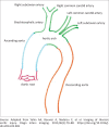
Traumatic aortic injury: Computed tomography angiography imaging and findings revisited in patients surviving major thoracic aorta injuries
- PMID: 33824749
- PMCID: PMC8008191
- DOI: 10.4102/sajr.v25i1.2044
Traumatic aortic injury: Computed tomography angiography imaging and findings revisited in patients surviving major thoracic aorta injuries
Abstract
Blunt chest trauma related acute thoracic aortic injury (TAI) is a life-threatening condition that requires prompt diagnosis and appropriate management because of high mortality. Computed tomography angiography (CTA) is the imaging of choice for evaluation of patients with major chest trauma findings suspicious of TAI on chest radiography. This case series describes the CTA findings in four high-velocity incident survivors with associated TAIs, discusses the injury type and treatment, and reviews the literature.
Keywords: TAI; computed tomography angiography; major chest trauma; thoracic aortic injuries; traumatic aortic injury.
© 2021. The Authors.
Conflict of interest statement
The authors declare that they have no financial or personal relationships that may have inappropriately influenced them in writing this article.
Figures


Similar articles
-
Cross-sectional imaging of thoracic traumatic aortic injury.Vasa. 2019 Jan;48(1):6-16. doi: 10.1024/0301-1526/a000741. Epub 2018 Sep 28. Vasa. 2019. PMID: 30264668 Review.
-
Thoracic aortic injury: how predictive is mechanism and is chest computed tomography a reliable screening tool? A prospective study of 1,561 patients.J Trauma. 2000 Apr;48(4):673-82; discussion 682-3. doi: 10.1097/00005373-200004000-00015. J Trauma. 2000. PMID: 10780601
-
The utility of intravascular ultrasound compared to angiography in the diagnosis of blunt traumatic aortic injury.J Vasc Surg. 2011 Mar;53(3):608-14. doi: 10.1016/j.jvs.2010.09.059. Epub 2010 Dec 3. J Vasc Surg. 2011. PMID: 21129901
-
To reduce routine computed tomographic angiography for thoracic aortic injury assessment in level II blunt trauma patients using three mediastinal signs on the initial chest radiograph: a preliminary report.Emerg Radiol. 2018 Aug;25(4):387-391. doi: 10.1007/s10140-018-1596-9. Epub 2018 Mar 13. Emerg Radiol. 2018. PMID: 29536276
-
Imaging of blunt aortic and great vessel trauma.J Thorac Imaging. 2000 Apr;15(2):97-103. doi: 10.1097/00005382-200004000-00004. J Thorac Imaging. 2000. PMID: 10798628 Review.
Cited by
-
Current updates in acute traumatic aortic injury: radiologic diagnosis and management.Clin Exp Emerg Med. 2022 Jun;9(2):73-83. doi: 10.15441/ceem.22.233. Epub 2022 Jun 30. Clin Exp Emerg Med. 2022. PMID: 35843607 Free PMC article.
-
Endovascular Aortic Repair in Traumatic Descending Thoracic Aortic Transection: A Case Report.Cureus. 2024 Sep 6;16(9):e68787. doi: 10.7759/cureus.68787. eCollection 2024 Sep. Cureus. 2024. PMID: 39371759 Free PMC article.
References
Publication types
LinkOut - more resources
Full Text Sources
Other Literature Sources
