Case Reports
. 2021 Mar 16:2021:6663519.
doi: 10.1155/2021/6663519. eCollection 2021.

From Neck Pain to Sarcoidosis: The Interesting Association

Affiliations
Case Reports

From Neck Pain to Sarcoidosis: The Interesting Association

Fadi Kharouf et al. Case Rep Rheumatol. .

Abstract

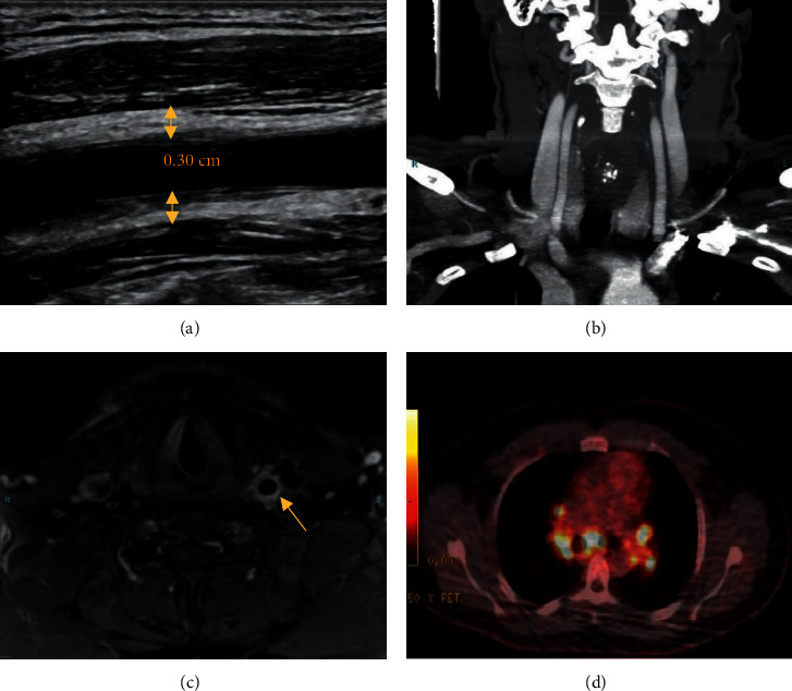
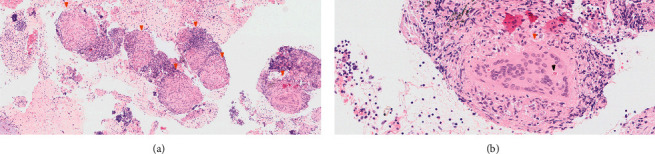
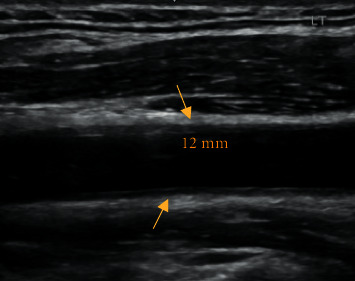
We report the case of a 31-year-old male patient, presenting to the emergency department (ED) with a 6-week history of left-sided lateral neck pain, along with a minor localized swelling. A few weeks after the beginning of his complaints, he contracted a mild coronavirus disease 2019 (COVID-19). Upon examination, his aches were defined as carotidynia; thus, proper radiologic evaluation was carried out. While ultrasound (US) and magnetic resonance imaging (MRI) scans showed evident signs of left common carotid (LCC) vasculitis, computed tomography angiography (CTA) and positron emission tomography-CT (PET-CT) scans revealed no vascular findings. Unexpected hypermetabolic hilar and mediastinal lymphadenopathy was found on PET-CT, necessitating lymph node biopsy. Pathology results displayed noncaseating granulomas. Besides, angiotensin-converting enzyme (ACE) levels in blood were high. Sarcoidosis, with concurrent LCC vasculitis, was diagnosed, and corticosteroid therapy was started. Shortly thereafter, remarkable recovery ensued.


Conflict of interest statement

The authors have no conflicts of interest to declare.

Figures

Figure 1
Figure 1
(a) Longitudinal ultrasound view showing LCC wall thickening (arrows). 0.30 cm relates to vessel wall thickness. (b) Coronal CTA view showing no LCC wall thickening. (c) Axial MRI view showing prominent LCC wall thickening. (d) PET/CT scan showing hypermetabolic mediastinal and hilar lymphadenopathy. Dimensions not to scale. CTA : computed tomography angiography; LCC : left common carotid; PET : positron emission tomography; MRI : magnetic resonance imaging.
Figure 2
Figure 2
(a) Numerous noncaseating sarcoid-like granulomas (orange arrowheads). (b) Well-formed granuloma (orange arrowheads) with a developing Schaumann body (black arrowhead).
Figure 3
Figure 3
Longitudinal ultrasound view showing a noticeable improvement in LCC wall thickening (arrows) after corticosteroid therapy, as compared to Figure 1(a). 0.12 cm relates to vessel wall thickness. Dimensions not to scale. LCC : left common carotid.

References

    1. Newman L. S., Rose C. S., Maier L. A. Sarcoidosis. New England Journal of Medicine. 1997;336(17):1224–1234. doi: 10.1056/NEJM199704243361706. - DOI - PubMed
    1. Villa-Forte, Hoffman G. Systemic vasculitis in sarcoidosis. In: Hoffman G. S., Weyand C. M., Langford C. A., Goronzy J. J., editors. Inflammatory Diseases of Blood Vessels. 2nd. Hoboken, NJ, USA: Blackwell Publishing Ltd; 2012. pp. 451–459. - DOI
    1. Fernandes S. R. M., Singsen B. H., Hoffman G. S. Sarcoidosis and systemic vasculitis. Seminars in Arthritis and Rheumatism. 2000;30(1):33–46. doi: 10.1053/sarh.2000.8364. - DOI - PubMed
    1. Weiler V., Redtenbacher S., Bancher C., Fischer M., Smolen J. Concurrence of sarcoidosis and aortitis: case report and review of the literature. Annals of the Rheumatic Diseases. 2000;59(11):850–853. doi: 10.1136/ard.59.11.850. - DOI - PMC - PubMed
    1. Dejaco C., Ramiro S., Duftner C., et al. EULAR recommendations for the use of imaging in large vessel vasculitis in clinical practice. Annals of the Rheumatic Diseases. 2018;77(5):636–643. doi: 10.1136/annrheumdis-2017-212649. - DOI - PubMed

Publication types

LinkOut - more resources