. 2021 Mar 20:2021:6697900.
doi: 10.1155/2021/6697900. eCollection 2021.

Evaluation of the PE Δ III-LC3-KDEL3 Chimeric Protein of Entamoeba histolytica- Lectin as a Vaccine Candidate against Amebic Liver Abscess

Affiliations

Evaluation of the PE Δ III-LC3-KDEL3 Chimeric Protein of Entamoeba histolytica- Lectin as a Vaccine Candidate against Amebic Liver Abscess

Sandra L Martínez-Hernández et al. J Immunol Res. .

Abstract

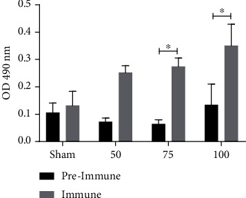
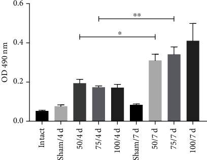
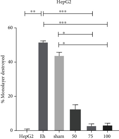
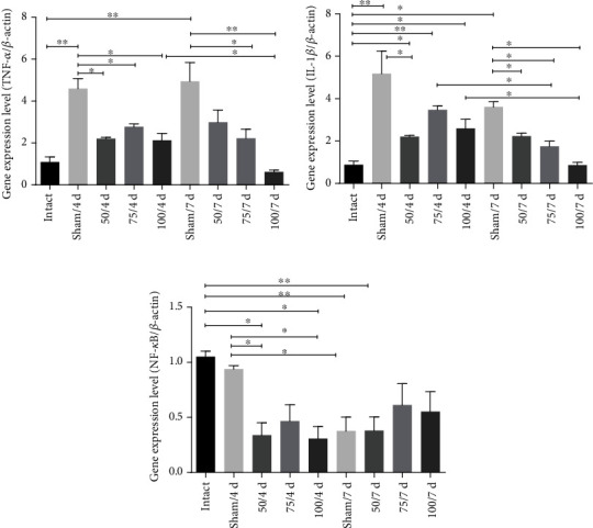
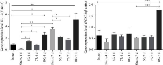
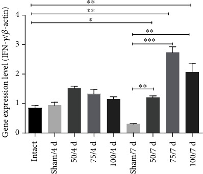
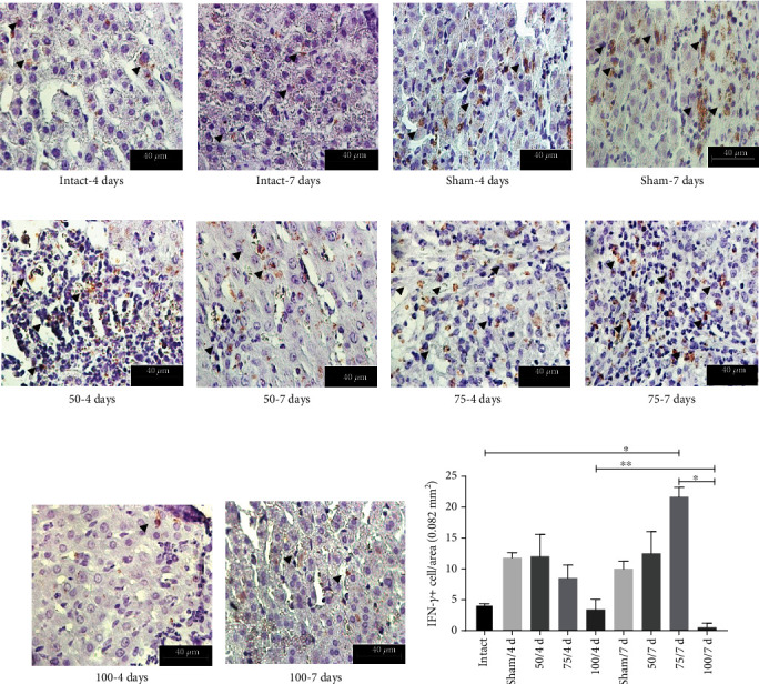
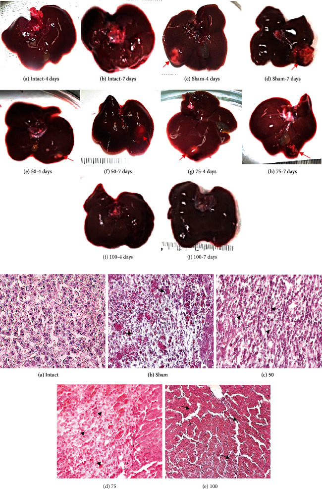
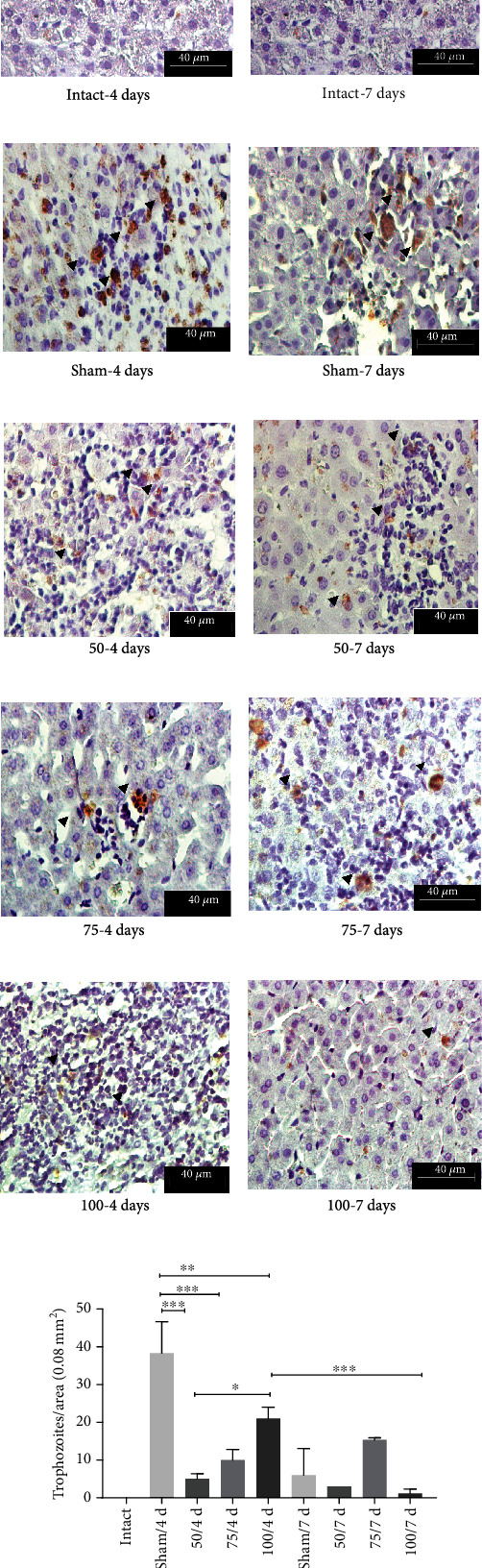
Entamoeba histolytica is an intestinal parasite that causes dysentery and amebic liver abscess. E. histolytica has the capability to invade host tissue by union of virulence factor Gal/GalNAc lectin; this molecule induces an adherence-inhibitory antibody response as well as to protect against amebic liver abscess (ALA). The present work showed the effect of the immunization with PEΔIII-LC3-KDEL3 recombinant protein. In vitro, this candidate vaccine inhibited adherence of E. histolytica trophozoites to HepG2 cell monolayer, avoiding the cytolysis, and in a hamster model, we observed a vaccine-induced protection against the damage to tissue liver and the inhibition of uncontrolled inflammation. PEΔIII-LC3-KDEL3 reduced the expression of TNF-α, IL-1β, and NF-κB in all immunized groups at 4- and 7-day postinfection. The levels of IL-10, FOXP3, and IFN-γ were elevated at 7 days. The immunohistochemistry assay confirmed this result, revealing an elevated quantity of +IFN-γ cells in the liver tissue. ALA formation in hamsters immunized was minimal, and few trophozoites were identified. Hence, immunization with PEΔIII-LC3-KDEL3 herein prevented invasive amebiasis, avoided an acute proinflammatory response, and activated a protective response within a short time. Finally, this recombinant protein induced an increase of serum IgG.


Conflict of interest statement

The authors declare that there is no conflict of interest regarding the publication of this paper.

Figures

Figure 1
Figure 1
The PEΔIII-LC3-KDEL3 vaccine induced IgG antibody production. Serum samples were evaluated by ELISA. Bars represent the mean ± SEM of three independent assays. Statistical analysis was performed with one-way ANOVA (p < 0.05).
Figure 2
Figure 2
The antibody levels were elevated in the animals receiving the PEΔIII-LC3-KDEL3 vaccine even in the postinfection period.
Figure 3
Figure 3
Inhibition of cytopathic effect of E. histolytica on HepG2 cells. Serum from the immunized groups inhibits destruction. Serum from sham animals generated a high percentage of destruction, similar to the positive control. HepG2 cells not exposed to E. histolytica represented the negative control. Data are expressed as the mean ± SEM of three independent assays, no parametric test Dunn's post hoc. p < 0.05; ∗∗p < 0.01; ∗∗∗p < 0.001.
Figure 4
Figure 4
The vaccine reduced the expression of the proinflammatory genes. (a, b) TNF-α and IL-1β in all immunized hamsters at 4- and 7-day postinfection were downregulated. (c) The NF-κB gene expression was also diminished in all immunized animals. Comparisons among groups: p < 0.05 and ∗∗p < 0.01.
Figure 5
Figure 5
The PEΔIII-LC3-KDEL3 vaccine upregulated IL-10 and FOXP3. (a, b) The 100 μg concentration of the vaccine induced a greater IL-10 and FOXP3 gene expression at 7-day postinfection. (b) Comparisons between groups: p < 0.05, ∗∗p < 0.01, and ∗∗∗p < 0.001.
Figure 6
Figure 6
The PEΔIII-LC3-KDEL3 vaccine promoted the expression of IFN-γ. The IFN-γ gene expression in the 50, 75, and 100 μg groups was only elevated at 7-day postinfection. Comparisons between groups: p < 0.05, ∗∗p < 0.01, and ∗∗∗p < 0.001.
Figure 7
Figure 7
Immunohistochemical analysis of +IFN-γ hepatic cells. Original magnification 400x. (a–j) Based on the immunohistochemistry assay, the quantity of +IFN-γ cells was determined in the liver tissue of hamsters (arrowhead). (k) The cells were abundant in 75 μg group at 7 days.
Figure 8
Figure 8
(a) Macroscopic analysis of amebic liver abscess formation. (A, B) No abscesses were detected in the liver of uninfected hamsters. (C, D) The liver of the sham group showed the characteristic ALA lesions. (E, F) The 50 μg group exhibited smaller lesions on day 7 than day 4 than sham. (G, H) A single small lesion is seen in the 75 μg group (arrows). (I, J) No lesion was detected in the liver of the 100 μg group. (b) Light microscopy of paraffin histological technique of amoebic liver abscess. Original magnification 400x. (A) In the intact hamsters, normal architecture was found in the liver tissue. (B, C) In two of the infected groups, sham and vaccinated with 50 μg, a necrotic area was observed in the liver parenchyma, accompanied by inflammatory infiltrate (arrows, asterisk). (D) In the 75 μg group, the parenchyma displayed no tissue necrosis (arrowhead). (E) In the 100 μg group, the parenchyma was similar to the intact animals, and there was no tissue necrosis or inflammation (arrows, cross).
Figure 9
Figure 9
Immunohistochemistry detection for E. histolytica trophozoites. Original magnification 400x. (c, d) Trophozoites and their fragments (e–j) can see by the light brown color (arrowheads). The number of trophozoites was lower as increment the vaccine dose (e–j). (k) Graphical expression of quantity of E. histolytica trophozoites per tissue area (p < 0.05, ∗∗p < 0.01, ∗∗∗p < 0.001).

Similar articles

Cited by

References

    1. Ximénez C., Cerritos R., Rojas L., et al. Human amebiasis: breaking the paradigm? International Journal of Environmental Research and Public Health. 2010;7(3):1105–1120. doi: 10.3390/ijerph7031105. - DOI - PMC - PubMed
    1. WHO. A moebiasis. Releve Epidemiologique. 1997;72(14):97–99. - PubMed
    1. Sack R. B., Haque R., Mondal D., Petri W. A., Kirkpatrick B. D. Attribution of malnutrition to cause-specific diarrheal illness: evidence from a prospective study of preschool children in Mirpur, Dhaka, Bangladesh. The American Journal of Tropical Medicine and Hygiene. 2009;80(5):824–826. doi: 10.4269/ajtmh.2009.80.824. - DOI - PMC - PubMed
    1. Haque R., Huston C. D., Hughes M., Houpt E., Petri W. A., Jr. Amoebiasis: review article. The New England Journal of Medicine. 2003;348(16):1565–1573. doi: 10.1056/NEJMra022710. - DOI - PubMed
    1. Ghosh S., Padalia J., Moonah S. Tissue destruction caused by Entamoeba histolytica parasite: cell death, inflammation, invasion, and the gut microbiome. Current Clinical Microbiology Reports. 2019;6(1):51–57. doi: 10.1007/s40588-019-0113-6. - DOI - PMC - PubMed

MeSH terms

LinkOut - more resources