Review
. 2021 Apr 5:6:26.
doi: 10.21037/tgh-2020-04. eCollection 2021.

Fibrocystic liver disease: novel concepts and translational perspectives

Affiliations
Review

Fibrocystic liver disease: novel concepts and translational perspectives

Alberto Lasagni et al. Transl Gastroenterol Hepatol. .

Abstract

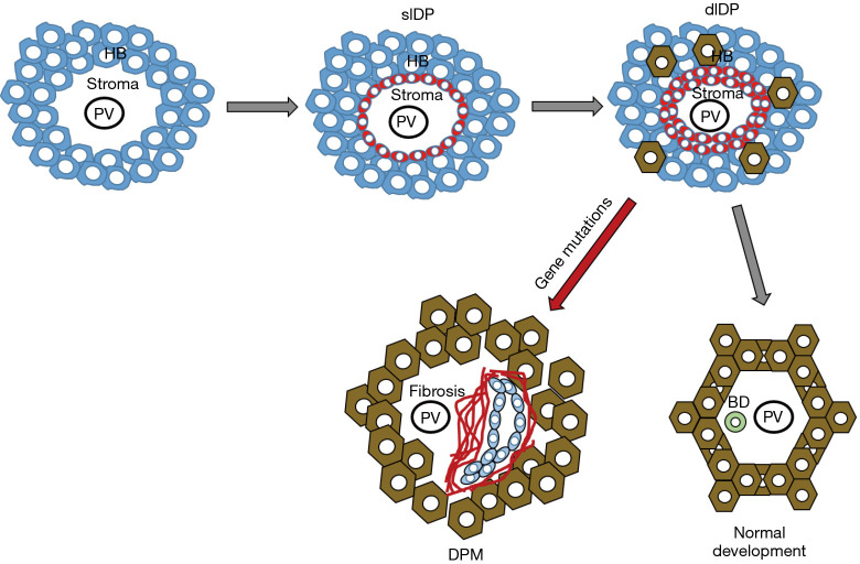
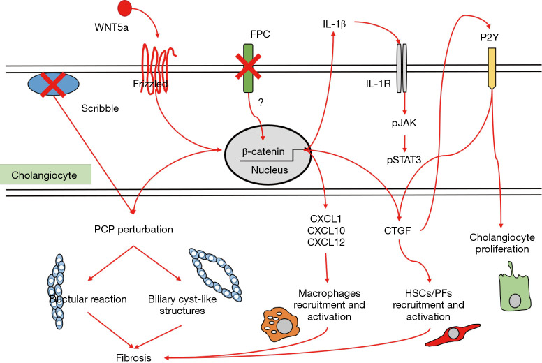
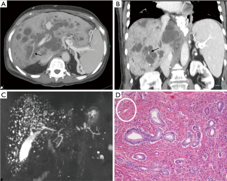
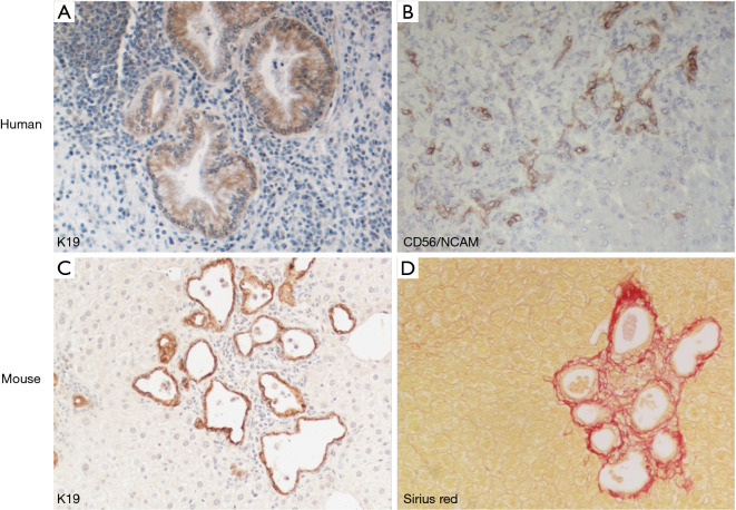
Fibrocystic liver diseases (FLDs) comprise a heterogeneous group of rare diseases of the biliary tree, having in common an abnormal development of the embryonic ductal plate caused by genetically-determined dysfunctions of proteins expressed in the primary cilia of cholangiocytes (and therefore grouped among the "ciliopathies"). The ductal dysgenesis may affect the biliary system at multiple levels, from the small intrahepatic bile ducts [congenital hepatic fibrosis (CHF)], to the larger intrahepatic bile ducts [Caroli disease (CD), or Caroli syndrome (CS), when CD coexists with CHF], leading to biliary microhamartomas and segmental bile duct dilations. Biliary changes are accompanied by progressive deposition of abundant peribiliary fibrosis. Peribiliary fibrosis and biliary cysts are the fundamental lesions of FLDs and are responsible for the main clinical manifestations, such as portal hypertension, recurrent cholangitis, cholestasis, sepsis and eventually cholangiocarcinoma. Furthermore, FLDs often associate with a spectrum of disorders affecting primarily the kidney. Among them, the autosomal recessive polycystic kidney disease (ARPKD) is the most frequent, and the renal function impairment is central in disease progression. CHF, CD/CS, and ARPKD are caused by a number of mutations in polycystic kidney hepatic disease 1 (PKHD1), a gene that encodes for fibrocystin/polyductin, a protein of unclear function, but supposedly involved in planar cell polarity and other fundamental cell functions. Targeted medical therapy is not available yet and thus the current treatment aims at controlling the complications. Interventional radiology or surgical treatments, including liver transplantation, are used in selected cases.

Keywords: Caroli disease (CD); Caroli syndrome (CS); Fibrocystic liver disease (FLD); biliary fibrosis; congenital hepatic fibrosis (CHF); polycystic kidney hepatic disease 1 (PKHD1).


Conflict of interest statement

Conflicts of Interest: All authors have completed the ICMJE uniform disclosure form (available at http://dx.doi.org/10.21037/tgh-2020-04). The series “Recent Advances in Rare Liver Diseases” was commissioned by the editorial office without any funding or sponsorship. LF and MS served as the unpaid Guest Editors of the series. LF serves as an unpaid editorial board member of Translational Gastroenterology and Hepatology from Sep 2018 to Aug 2020. MS is a member of the advisory board of Esiai/Merk, Bayer, and Engitix, during the conduct of the study. The authors have no other conflicts of interest to declare.

Figures

Figure 1
Figure 1
Fetal development of intrahepatic bile duct epithelium is a highly coordinated process involving several different cell types. Initially, the hepatoblasts (HB) aligning along the nascent portal tract centered around the portal vein (PV) branches are primed to gain biliary-like characteristics and transdifferentiate into single layer ductal plate (slDP, red cells), the primordial structure of the bile duct, that further progresses to double layer ductal plate (dlDP). In normal conditions, dlDP migrate into the portal mesenchyme to become incorporated as fully differentiated bile ducts (BD). When this maturation process is perturbed, as by genetic defects, DPM develop at the margins of the portal tract, and are often associated with the nearby deposition of fibrotic tissue.
Figure 2
Figure 2
Main molecular mechanisms contributing to cyst development, which act through deregulation of planar cell polarity, stimulation of cholangiocyte proliferation and activation of inflammatory cells and myofibroblasts in the peribiliary area. They involve a complex interplay of numerous cytokines, chemokines, growth factors and morphogens, regulated by β-catenin and pJAK/STAT3 signaling. Of note, perturbation of planar cell polarity may lead to ductular reaction or cyst-like structure formation, accompanied by progressive peribiliary fibrosis. See text for details. FPC, fibrocystin/polyductin; PCP, planar cell polarity; CTGF, connective tissue growth factor; HSCs, hepatic stellate cells; PFs, portal fibroblasts.
Figure 3
Figure 3
Radiological and histological findings in CD. Axial (A) and coronal (B) reconstruction of 83-year-old woman using contrast-enhanced CT. Images outline the diffuse cystic changes of the intrahepatic bile ducts with the typical “central dot sign” (arrows). MR coronal view (C) of a 68-year-old man showing the typical “starry sky” appearance consistent with the presence of multiple microhamartomas. H&E histological staining (×10) (D) show biliary microhamartomas surrounded by a dense stromal reaction composed by fibrotic bundles and inflammatory cells. On the upper left corner (white circle), hepatic artery and portal vein branches are embedded in a dense fibrotic area (histological lesion equivalent of the radiological “central dot sign”).
Figure 4
Figure 4
Liver phenotype in FLD, in human and experimental mouse model. Dysgenetic bile ducts immunoreactive for K19 develop a cyst-like configuration in enlarged portal tracts containing an abundant inflammatory cell infiltrate (A, ×20). They are accompanied by ductal plate remnants expressing CD56/NCAM (neural cell adhesion molecule) mostly localized at the portal tract margins (B, ×10). These features are phenocopied by the Pkhd1del4/del4 mouse, where microhamartomas and biliary cysts expressing K19 (C, ×10) are tightly surrounded by extensive peribiliary fibrosis confined to the portal area, as shown by histochemical staining for Sirius Red (D, ×10).

References

    1. Desmet VJ. Pathogenesis of Ductal Plate Abnormalities. Mayo Clin Proc 1998;73:80-9. 10.1016/S0025-6196(11)63624-0 - DOI - PubMed
    1. Gunay-Aygun M. Liver and kidney disease in ciliopathies. Am J Med Genet C Semin Med Genet 2009;151C:296-306. 10.1002/ajmg.c.30225 - DOI - PMC - PubMed
    1. Gascue C, Katsanis N, Badano JL. Cystic diseases of the kidney: ciliary dysfunction and cystogenic mechanisms. Pediatr Nephrol 2011;26:1181-95. 10.1007/s00467-010-1697-5 - DOI - PMC - PubMed
    1. Avidor-Reiss T, Maer AM, Koundakjian E, et al. Decoding cilia function: defining specialized genes required for compartmentalized cilia biogenesis. Cell 2004;117:527-39. 10.1016/S0092-8674(04)00412-X - DOI - PubMed
    1. Fliegauf M, Benzing T, Omran H. When cilia go bad: cilia defects and ciliopathies. Nat Rev Mol Cell Biol 2007;8:880-93. 10.1038/nrm2278 - DOI - PubMed

LinkOut - more resources