Review
. 2021 Aug;385(2):405-422.
doi: 10.1007/s00441-021-03429-4. Epub 2021 Apr 6.

Perspectives in membranous nephropathy

Affiliations
Review

Perspectives in membranous nephropathy

Nicola M Tomas et al. Cell Tissue Res. 2021 Aug.

Abstract

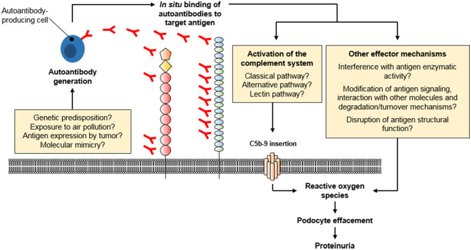
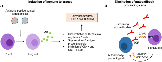
The identification of the phospholipase A2 receptor 1 (PLA2R) and thrombospondin type-1 domain-containing protein 7A (THSD7A) as podocyte antigens in adult patients with membranous nephropathy (MN) has strongly impacted both experimental and clinical research on this disease. Evidence has been furnished that podocyte-directed autoantibodies can cause MN, and novel PLA2R- and THSD7A-specific animal models have been developed. Today, measurement of serum autoantibody levels and staining of kidney biopsies for the target antigens guides MN diagnosis and treatment worldwide. Additionally, anti-PLA2R antibodies have been proven to be valuable prognostic biomarkers in MN. Despite these impressive advances, a variety of questions regarding the disease pathomechanisms, clinical use of antibody measurement, and future treatments remain unanswered. In this review, we will outline recent advances made in the field of MN and discuss open questions and perspectives with a focus on novel antigen identification, mechanisms of podocyte injury, clinical use of antibody measurement to guide diagnosis and treatment, and the potential of innovative, pathogenesis-based treatment strategies.

Keywords: Membranous nephropathy; PLA2R; THSD7A.


Conflict of interest statement

The authors declare that there is no conflict of interest.

Figures

Fig. 1
Fig. 1
Morphological findings in a patient with membranous nephropathy. (a) Periodic acid Schiff (PAS) staining demonstrates prominent glomerular capillary walls (black arrows) as a sign of a thickened glomerular basement membrane (GBM). (b, c) Immunohistochemical staining for IgG (b) and complement component C3 (c) shows strong positivity (red), indicating deposition of (auto)antibodies and C3. (d) Electron microscopy demonstrates a thickened GBM with electron dense deposits accentuated at the outer (subepithelial) aspect of the GBM (light blue arrows) and effacement of podocyte foot processes (light blue asterisks). Images kindly provided by Thorsten Wiech, University Medical Center Hamburg-Eppendorf
Fig. 2
Fig. 2
Molecular architecture of MN antigens/biomarkers. (a) PLA2R is a 180 kDa plasma membrane protein consisting of a cysteine-rich (CysR) domain, a fibronectin type II (FnII) domain, eight c-type lectin domains (CTLDs), one transmembrane region and a short cytoplasmic tail. (b) THSD7A is a 250 kDa plasma membrane protein consisting of twenty-one thrombospondin repeat (TSR) domains, which can be further classified as thrombospondin (THBS1)-like (blue) or F-spondin/C6-like (green), one transmembrane region and a short cytoplasmic tail. (c) EXT1 and EXT2 are proteins expressed in the endoplasmic reticulum membrane with a short cytoplasmic tail, one transmembrane region, short stem region and a long luminal tail. EXT1 contains a luminal catalytic domain. (d) NELL1 is a 90 kDa cytosolic protein containing a THBS1-like domain, one coiled-coil domain, 4 von Willebrandt (VW)-type domains and six epidermal growth factor-like (EGF-like) domains. (e) SEMA3B is a 80 kDa secreted protein containing a Sema domain, a plexin-semaphorinintegrin (PSI) domain, an immunoglobulin (IgG) domain and a basic domain
Fig. 3
Fig. 3
Molecular pathogenesis of MN. The initial step in the development of MN is the generation of autoantibodies. The initiation triggers of autoantibody generation have not been resolved, but several mechanisms (or a combination thereof) have been proposed: carriage of certain risk alleles, long-time exposure to air pollution, antigen expression by malignant tumors and molecular similarities of the antigens with, e.g., pathogens. Autoantibodies then bind to their target antigens on podocytes, which leads to the local activation of the complement system. Additional mechanisms of podocyte injury, such as disturbance of antigen function and signaling, have been suggested to play a role in MN pathogenesis, yet this needs further clarification
Fig. 4
Fig. 4
Innovative strategies to treat MN. (a) Antigen-specific tolerance can be induced using nanoparticles coated with antigen peptides corresponding to the T cell epitopes. Such nanoparticles can induce the differentiation of proinflammatory TH1 cells to regulatory T (Treg) cells, which produce interleukin-10 (IL-10). IL-10 induces the differentiation of B cells to regulatory B cells, the suppression of antigen-presenting cells and the inhibition of different T cell populations, collectively decreasing the immune response against the respective specific antigen. (b) Autoantibody-producing cells can be eliminated based on the specificity of the B cell receptor (BCR). The BCR is a membrane-bound antibody, corresponding to the antibody that is produced by this exact cell. T and NK cells can be transduced with chimeric autoantibody receptors, consisting of antigen fragments coupled to a transmembrane and several intracellular signaling domains. Depicted is a CAAR containing the first three domains of PLA2R, the cysteine-rich domain, the fibronectin type II domain, and the first c-type lectin domain. Binding of the CAAR-transduced cell to the BCR induces signaling events leading to the release of granzyme and perforins, which can efficiently eliminate the nearby autoantibody-producing cell

References

    1. Alto LT, Terman JR. Semaphorins and their signaling mechanisms. Methods Mol Biol. 2017;1493:1–25. doi: 10.1007/978-1-4939-6448-2_1. - DOI - PMC - PubMed
    1. Augert A, Payre C, de Launoit Y, Gil J, Lambeau G, Bernard D. The M-type receptor PLA2R regulates senescence through the p53 pathway. EMBO Rep. 2009;10(3):271–277. doi: 10.1038/embor.2008.255. - DOI - PMC - PubMed
    1. Baker PJ, Ochi RF, Schulze M, Johnson RJ, Campbell C, Couser WG. Depletion of C6 prevents development of proteinuria in experimental membranous nephropathy in rats. Am J Pathol. 1989;135(1):185–194. - PMC - PubMed
    1. Bally S, Debiec H, Ponard D, Dijoud F, Rendu J, Faure J, Ronco P, Dumestre-Perard C. Phospholipase A2 receptor-related membranous nephropathy and mannan-binding lectin deficiency. J Am Soc Nephrol. 2016;27(12):3539–3544. doi: 10.1681/ASN.2015101155. - DOI - PMC - PubMed
    1. Bech AP, Hofstra JM, Brenchley PE, Wetzels JF. Association of anti-PLA(2)R antibodies with outcomes after immunosuppressive therapy in idiopathic membranous nephropathy. Clin J Am Soc Nephrol. 2014;9(8):1386–1392. doi: 10.2215/CJN.10471013. - DOI - PMC - PubMed

MeSH terms