Review
. 2021 May 1;125(5):1899-1919.
doi: 10.1152/jn.00017.2021. Epub 2021 Apr 7.

Neuronal mechanisms underlying opioid-induced respiratory depression: our current understanding

Affiliations
Review

Neuronal mechanisms underlying opioid-induced respiratory depression: our current understanding

Jan-Marino Ramirez et al. J Neurophysiol. .

Abstract

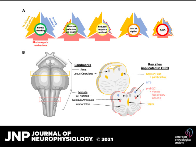
Opioid-induced respiratory depression (OIRD) represents the primary cause of death associated with therapeutic and recreational opioid use. Within the United States, the rate of death from opioid abuse since the early 1990s has grown disproportionally, prompting the classification as a nationwide "epidemic." Since this time, we have begun to unravel many fundamental cellular and systems-level mechanisms associated with opioid-related death. However, factors such as individual vulnerability, neuromodulatory compensation, and redundancy of opioid effects across central and peripheral nervous systems have created a barrier to a concise, integrative view of OIRD. Within this review, we bring together multiple perspectives in the field of OIRD to create an overarching viewpoint of what we know, and where we view this essential topic of research going forward into the future.

Keywords: OIRD; breathing; neuromodulation; opioid; respiration.


Conflict of interest statement

No conflicts of interest, financial or otherwise, are declared by the authors.

Figures

None
Graphical abstract
Figure 1.
Figure 1.
A: normal breathing requires the integration of rhythmogenic, modulatory, and sensory feedback mechanisms. Opioid overdose can suppress all of these important mechanisms of respiratory control, leading to OIRD. B: human brainstem schematic illustrating key sites in the pons and medulla that mediate OIRD. Ventral view is shown on the left, and transverse sections are shown on the right. Colors in B correspond to mechanisms depicted in A. NTS, nucleus tractus solitarius; OIRD, opioid-induced respiratory depression; preBOTc, preBӧtzinger complex.
Figure 2.
Figure 2.
Examples of neurons with altered discharge identity or discharge rate after codeine administration from several animals. Several of the neurons shown became breathing modulated (examples 1–3). In others, the time of maximum discharge rate during the respiratory cycle changed (4 and 5). Discharge rates of most of the neurons shown increased (1, 2, and 7) or decreased (4 and 5) after codeine. Cycle-triggered histograms of neuronal activity (colors) overlying phrenic discharge (gray). E-P, expiratory phasic; E-T, expiratory tonic; I-T, inspiratory tonic; n, neuron; NBM, nonbreathing modulated. Anesthetized, paralyzed, and artificially ventilated cats. Codeine (1–3,000 µg/kg) was administered via the vertebral artery.
Figure 3.
Figure 3.
Schematic illustrating the canonical MOR signaling pathways via Gi/o-protein coupling. Gβγ can directly activate GIRK currents and directly inhibit voltage-gated Ca2+ currents. Gαi inhibits adenylyl cyclase, reducing the intracellular concentration of cyclic AMP. Reduced cAMP concentrations inhibit voltage-activated cation currents, such as HCN and can also indirectly influence the activity of numerous downstream effectors through inhibition of PKA. The predicted effects of these mechanisms on preBötC network function and the inspiratory rhythm depend on their somatic and/or synaptic sites of action. Predicted effects are color coded to the canonical pathways outlined above. GIRK, G-protein-activated inward rectifier K+ channels; MOR, mu-opioid receptor; preBOTc, preBӧtzinger complex.
Figure 4.
Figure 4.
A: schematic illustrating the proposed effect of opioids at excitatory synapses on rhythmogenic Dbx1 neurons. In response to MOR activation, Gβγ-mediated inhibition of voltage-gated Ca2+ currents reduces the probability of vesicular release in response to bouts of action potentials that occur during inspiratory bursts. B: PreBötC slices from mice with a heterozygous deletion of CaV 2.1 encoding P/Q-type Ca2+ channels are ∼10-fold more sensitive to the MOR agonist DAMGO than WT controls (data shown as median ± interquartile range). C: representative voltage clamp recordings of mEPSCs from a Dbx1 neuron following blockade of action potentials with TTX (1 µM). The frequency of mEPSCs is reduced in the presence of DAMGO (100 nM), and subsequent blockade KCNQ channels with XE991 (20 µM) restores mEPSC frequency to baseline levels. Data adapted with permission from Wei and Ramirez (20). KO, knockout; mEPSCs, miniature excitatory postsynaptic currents; MOR, mu-opioid receptor; P, probability; preBOTc, preBӧtzinger complex; WT, wild type.
Figure 5.
Figure 5.
Opioid modulation of pontine networks. A: whole cell voltage-clamp recording from a KF neuron in brain slice illustrating activation of GIRK current by opioid agonist Met-enkephalin. Modified from Levitt et al. (184). B: schematic of the in situ working heart-brainstem preparation of the rat. On the right, phrenic bursts indicating inspiratory activity during baseline, following systemic fentanyl administration, and following systemic fentanyl with opioid antagonist, CTAP microinjection into the KF. Antagonism of KF MORs eliminates rate depression by fentanyl; however, the apneustic pattern characteristic of opioid effects persists. Modified from Saunders and Levitt, 2020 (211). C: schematic of in vivo plethysmograph. Graph on the top right demonstrates significant attenuation of morphine-induced respiratory rate depression in vivo following bilateral MOR deletion from KF neurons compared to control animals. Below, example time course of average respiratory rate following saline and morphine (10 mg/kg, i.p.) in control mice (left) vs. mice following MOR deletion from KF neurons (right). Modified from Varga et al. (212). GFP, green fluorescent protein; GIRK, G-protein-activated inward rectifier K+ channels; KF, Kölliker-Fuse; MOR, mu-opioid receptor. *,**,***Statistically significant (P < 0.05) in order of least to most significant.
Figure 6.
Figure 6.
Examples of how tapered spectrograms reveal that administering saline (A), morphine, (B), buprenorphine (C), or fentanyl (D) differentially changes cortical EEG power and frequency in mice. E illustrates how EEG power can be quantified to show opioid-specific alterations. These spectrograms demonstrate the importance of understanding nuanced effects of opioids on cortical excitability. Modified from O’Brien et al. (282).
Figure 7.
Figure 7.
An example of how breathing in mice is depressed by direct administration of fentanyl into the prefrontal cortex A: adequate respiratory control includes the ability to vary breathing in response to environmental challenges. B shows how Poincaré analyses can be used to visualize the decrease in breathing variability caused by delivering fentanyl into mouse prefrontal cortex. Modified from Zhang et al. 2019 (284). PFC, prefrontal cortex. *P < 0.05.

References

    1. Hedegaard H, Miniño AM, Warner M. Drug overdose deaths in the United States, 1999–2018. Hyattsville, MD: National Center for Health Statistics, 2020.
    1. National Academies of Sciences Engineering, and Medicine; Health and Medicine Division; Board on Health Sciences Policy; Committee on Pain Management and Regulatory Strategies to Address Prescription Opioid Abuse. Pain Management and the Opioid Epidemic: Balancing Societal and Individual Benefits and Risks of Prescription Opioid Use, edited byPhillips JK, Ford MA, Bonnie RJ.. Washington, DC: National Academic Press, 2017. - PubMed
    1. Rudd RA, Seth P, David F, Scholl L. Increases in drug and opioid-involved overdose deaths—United States, 2010–2015. MMWR Morb Mortal Wkly Rep 65: 1445–1452, 2016. doi:10.15585/mmwr.mm655051e1. - DOI - PubMed
    1. Scholl L, Seth P, Kariisa M, Wilson N, Baldwin G. Drug and opioid-involved overdose deaths—United States, 2013–2017. MMWR Morb Mortal Wkly Rep 67: 1419–1427, 2018. doi:10.15585/mmwr.mm675152e1. - DOI - PMC - PubMed
    1. Dahan A, Overdyk F, Smith T, Aarts L, Niesters M. Pharmacovigilance: a review of opioid-induced respiratory depression in chronic pain patients. Pain Physician 16: E85–E94, 2013. - PubMed

Publication types

MeSH terms

Substances