. 2021 Apr 6;35(1):108962.
doi: 10.1016/j.celrep.2021.108962.

The mechanistic basis of protection by non-neutralizing anti-alphavirus antibodies

Affiliations

The mechanistic basis of protection by non-neutralizing anti-alphavirus antibodies

James T Earnest et al. Cell Rep. .

Abstract

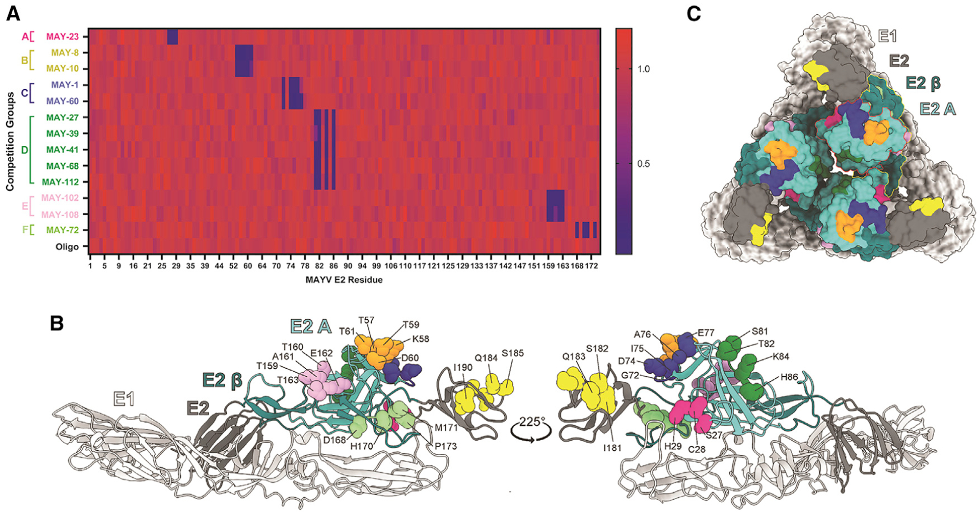
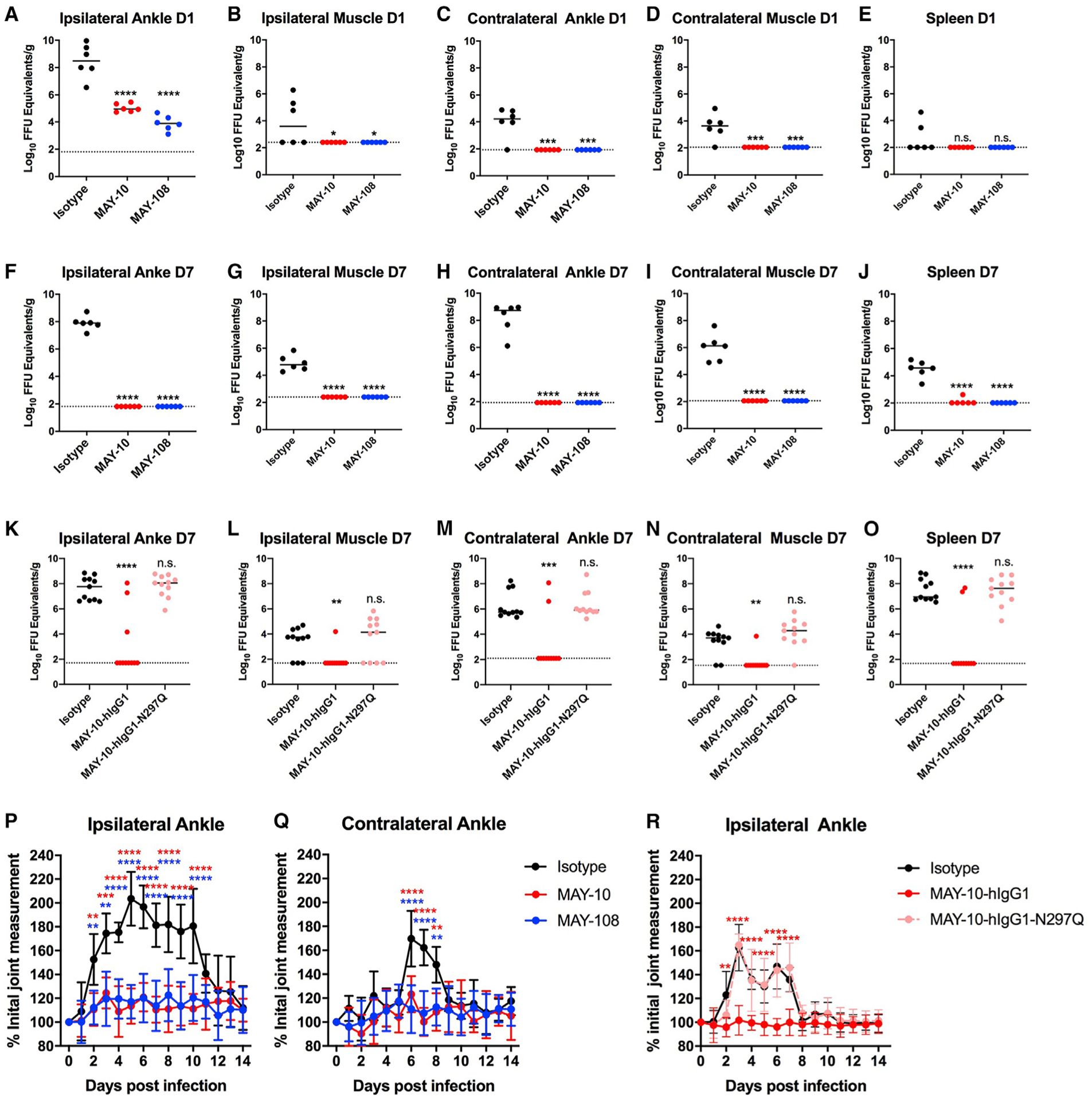
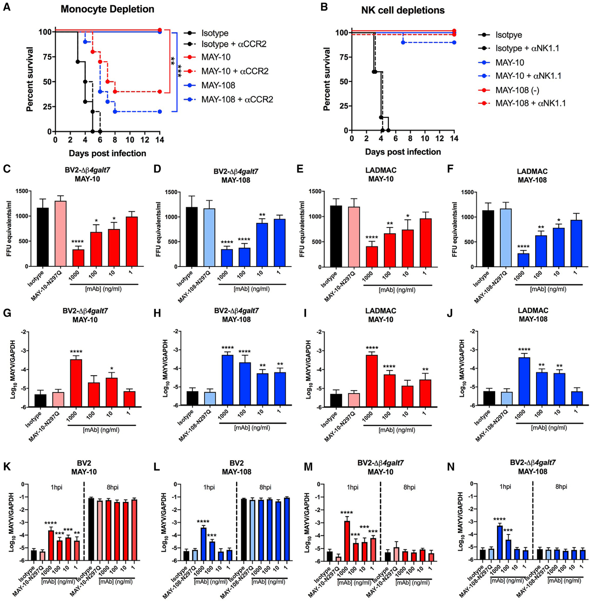
Although neutralizing monoclonal antibodies (mAbs) against epitopes within the alphavirus E2 protein can protect against infection, the functional significance of non-neutralizing mAbs is poorly understood. Here, we evaluate the activity of 13 non-neutralizing mAbs against Mayaro virus (MAYV), an emerging arthritogenic alphavirus. These mAbs bind to the MAYV virion and surface of infected cells but fail to neutralize infection in cell culture. Mapping studies identify six mAb binding groups that localize to discrete epitopes within or adjacent to the A domain of the E2 glycoprotein. Remarkably, passive transfer of non-neutralizing mAbs protects against MAYV infection and disease in mice, and their efficacy requires Fc effector functions. Monocytes mediate the protection of non-neutralizing mAbs in vivo, as Fcγ-receptor-expressing myeloid cells facilitate the binding, uptake, and clearance of MAYV without antibody-dependent enhancement of infection. Humoral protection against alphaviruses likely reflects contributions from non-neutralizing antibodies through Fc-dependent mechanisms that accelerate viral clearance.

Keywords: Fc effector; alphavirus; antibody; epitope; mapping; monocytes; neutralization; pathogenesis; protection.


Conflict of interest statement

Declaration of interests M.S.D. is a consultant for Inbios, Vir Biotechnology, NGM Biopharmaceuticals, and the Carnival Corporation and on the Scientific Advisory Board of Moderna and Immunome. The Diamond laboratory has received sponsored research agreements from Moderna, Vir Biotechnology, and Emergent BioSolutions.

Figures

Figure 1.
Figure 1.. Binding of non-neutralizing anti-MAYV mAbs to virions and recombinant E2 protein
(A and B) Anti-MAYV mAbs were tested for neutralization of MAYV on Vero (A) and C2C12 myoblast (B) cells. Serial dilutions of the indicated mAbs were incubated with 102 FFU of MAYV-BeH407 and then added to the indicated cells. Viral foci are plotted relative to a no mAb control. The neutralizing mAb MAY-117 was used as a positive control, and an irrelevant mIgG2c mAb was used as a negative isotype control (mean and SD of two experiments performed in triplicate). (C and D) Binding to MAYV virions (C) or recombinant MAYV E2 protein (D) by ELISA. Virions were captured with a humanized mAb to MAYV, and recombinant MAYV E2 protein was bound directly to microtiter plates. Bound murine mAbs were detected with an horseradish peroxidase (HRP)-conjugated secondary antibody. Data are expressed as OD values relative to the 10-μg/ml sample (mean and SD of two experiments performed in triplicate). (E) MAYV mAbs were competed for binding to MAYV (strain BeH407) by ELISA. Virus was captured on plates using a humanized anti-MAYV mAb. Captured virion was incubated with 10 μg/ml of the indicated mAb (first antibody). Antibody-virus complexes were incubated with 10 ng/ml of the indicated mAb labeled with biotin (second antibody). Binding was detected using streptavidin HRP and is indicated by color from high (red) to low (blue). Data are presented relative to a control with no first antibody and are representative of two experiments.
Figure 2.
Figure 2.. Mapping of mAbs to MAYV E2 protein
(A) Heatmap of relative binding of anti-MAYV mAbs to MAYV-E2 A domain mutants. 293T cells were transfected with a C-E3-E2–6K-E1 plasmid containing alanine mutations in the A domain of E2. Binding of the indicated mAbs were measured by flow cytometry; the full dataset is shown in Table S1 and Figure S2. Relative binding compared to an oligoclonal control is indicated by color from high (red) to low (blue). (B and C) Residues required for mAb engagement are depicted as balls and sticks on a ribbon diagram of the predicted structure of MAYV E2-E1 monomer generated using Phyre2 (B) and are highlighted on the monomers arranged as a trimeric spike (C). The E1 and E2 glycoproteins are light and dark gray, respectively. Within E2, domain A is cyan, the β-ribbon is dark cyan. In the surface representation (C), the A domain and β-ribbon regions are outlined in red and yellow, respectively. Competition groups are color coded as follows: group A, dark pink (residues 27–29); group B, orange (57–61); group C, blue (72–77); group D, dark green (81–86); group E, lavender (159–163); group F, light green (168–173); and the anti-B domain control mAb MAY-117, bright yellow (181–186). See also Table S1 and Figure S2.
Figure 3.
Figure 3.. Antibody protection against lethal MAYV challenge
Four-week-old C57BL/6J female mice were treated with 100 μg of anti-Ifnar1 mAb 1 day before subcutaneous inoculation of MAYV-BeH407. (A and B) A single 100-μg dose of anti-MAYV mAbs was administered by intraperitoneal injection 1 day before virus inoculation. The mAbs exhibited a range of activity with some showing >50% protection (A) and others <50% (B). Data are from two experiments. (C and D) Combination therapy of an anti-MAYV E2 B domain mAb (MAY-134) and anti-MAYV E2 A domain mAbs. C57BL6/J mice were treated with 100 μg of anti-Ifnar1 mAb 1 day before subcutaneous virus inoculation. (C) Two days after infection, mice were treated with 200 μg of MAY-10 or MAY-134 or 100 μg each of MAY-10 and MAY-134. (D) Two days after infection, mice were treated with 200 μg of MAY-108 or MAY-134 or 100 μg each of MAY-108 and MAY-134 (two experiments, n = 8; *p < 0.05; **p < 0.01; ***p < 0.001; ****p < 0.0001; log-rank test with Bonferroni correction compared to isotype control mAb). See also Figure S3.
Figure 4.
Figure 4.. Protection by non-neutralizing mAbs is Fc dependent
(A) Four-week-old C57BL/6J FcγR−/− male and female mice were administered 100 μg of MAY-10 or MAY-108 1 day before subcutaneous inoculation of MAYV-BeH407 (two experiments, n = 6). (B) Isotype-switched mAb binding to recombinant murine FcγRI, FcγRIII, and FcγRIV. MAY-10 or MAY-108 of the indicated isotype were added to plates coated with FcγRs. Binding data: two-way ANOVA with Sidak’s post-test, compared to mIgG2c isotype mAb. (C–F) MAY-10 (C) and MAY-108 (E) were isotype switched from murine IgG2c to murine IgG1, human IgG1, or human IgG1-N297Q. Each antibody variant was tested for binding to captured MAYV by ELISA. For protection studies (D and F), 100 μg of the indicated mAb was administered to 4-week-old C57BL/6J mice 1 day before subcutaneous inoculation with MAYV (two experiments, n = 6; log-rank test with Bonferroni correction compared to isotype control mAb). *p < 0.05; **p < 0.01; ***p < 0.001; ****p < 0.0001.
Figure 5.
Figure 5.. Antibodies clear MAYV, prevent viral dissemination, and protect against musculoskeletal disease
(A–J) Tissue titers of MAYV at 1 (A–E) or 7 (F–J) dpi. Four-week-old C57BL/6J mice treated with 100 μg of MAY-10, MAY-108, or an isotype control mAb 1 day before subcutaneous inoculation with MAYV-BeH407. At the indicated days, the ipsilateral ankle (A and F), ipsilateral calf muscle (B and G), contralateral ankle (C and H), contralateral calf muscle (D and I), and spleen (E and J) were harvested, and viral RNA was measured (two experiments; n = 6, one-way ANOVA with Dunnett’s post-test). (K–O) Tissue titers of mice treated with 100 μg of MAY-10-hIgG1 or MAY-hIgG1-N297Q 1 day before infection. MAYV titers in the ipsilateral ankle (K) and calf muscle (L), the contralateral ankle (M) and calf muscle (N), and the spleen (O) were measured at 7 dpi (three experiments; n = 11, one-way ANOVA with Dunnett’s post-test). (P–R) Four-week-old C57BL/6J mice were treated with MAY-10 or MAY-108 and infected as described above. Ipsilateral (P) and contralateral (Q) ankle joints were measured using digital calipers. (R) Ipsilateral ankle swelling was measured in mice treated with MAY-10-hIgG1 or MAY-10-hIgG1-N297Q (mean and SD of two experiments; n = 10, two-way ANOVA with Tukey’s post-test). *p < 0.05; **p < 0.01; ***p < 0.001; ****p < 0.0001. See also Figure S4.
Figure 6.
Figure 6.. Protection by non-neutralizing mAbs depends on monocytes
(A) Four-week-old C57BL6/J mice were treated with 100 μg of anti-Ifnar1 mAb and 100 μg of MAY-10, MAY-108, or isotype control mAb 1 day before infection with MAYV-BeH407. Indicated mice also were treated with 25 μg of an anti-CCR2 mAb at 1 day before infection and every other day after (two experiments, n = 10; log-rank test with Bonferroni correction). (B) NK cells were depleted by treating mice with 200 μg mAb of anti-NK1.1 mAb 1 day before infection and every other day after. Mice were treated with anti-Ifnar1 mAb and MAY-10, MAY-108, or isotype control mAb as above (two experiments, n = 10; log-rank test with Bonferroni correction). (C–J) Antibody-mediated binding of hIgG1 variants of MAYV to BV2-Δβ4galt7 (C, D, G, and H) or LADMAC (E, F, I, and J) cells. Virus binding to cells was measured indirectly by the depletion of MAYV from supernatants (C–F) or by direct binding and/or internalization of target cells (G–J). For measuring supernatants, MAYV was incubated with the indicated amount of hIgG1 variants of MAY-10 (C and E) or MAY-108 (D and F) for 1 h at 37°C before adding to the indicated cell for 30 min at 37°C. Viral RNA from supernatants was measured. Isotype-matched antibodies and hIgG1-N297Q mAb variants served as negative controls. To measure virus binding and internalization, BV2-Δβ4galt7 or LADMAC cells were inoculated with MAYV that had been pre-incubated with MAY-10 (G and I) or MAY-108 (H and J). After incubating for 30 min at 37°C, cells were washed with PBS and lysed, and viral RNA was measured. (K–N) Antibody-dependent infection assays. Serial dilutions of hIgG1 mAbs were pre-incubated with MAYV before being added to permissive BV2 cells (K and L) or non-permissive BV2-Δβ4galt7 cells (M and N). Binding and internalization into cells was measured as above (G–H) at 1 or 8 hpi after removal of unbound virus (mean and SD of three experiments performed in triplicate; one-way ANOVA with Dunnett’s post-test compared to the isotype mAb control). *p < 0.05; **p < 0.01; ***p < 0.001; ****p < 0.0001. See also Figures S5–S8.

References

    1. Azevedo RS, Silva EV, Carvalho VL, Rodrigues SG, Nunes-Neto JP, Monteiro H, Peixoto VS, Chiang JO, Nunes MR, and Vasconcelos PF (2009). Mayaro fever virus, Brazilian Amazon. Emerg. Infect. Dis 15, 1830–1832. - PMC - PubMed
    1. Bournazos S, DiLillo DJ, and Ravetch JV (2015). The role of Fc-FcγR interactions in IgG-mediated microbial neutralization. J. Exp. Med 212, 1361–1369. - PMC - PubMed
    1. Bournazos S, Corti D, Virgin HW, and Ravetch JV (2020a). Fc-optimized antibodies elicit CD8 immunity to viral respiratory infection. Nature 588, 485–490. - PMC - PubMed
    1. Bournazos S, Gupta A, and Ravetch JV (2020b). The role of IgG Fc receptors in antibody-dependent enhancement. Nat. Rev. Immunol 20, 633–643. - PMC - PubMed
    1. Brandt WE, McCown JM, Gentry MK, and Russell PK (1982). Infection enhancement of dengue type 2 virus in the U-937 human monocyte cell line by antibodies to flavivirus cross-reactive determinants. Infect. Immun 36, 1036–1041. - PMC - PubMed

Publication types

MeSH terms

LinkOut - more resources