. 2021 May 19;41(20):4367-4377.
doi: 10.1523/JNEUROSCI.2667-20.2021. Epub 2021 Apr 7.

Increased Excitatory Synaptic Transmission Associated with Adult Seizure Vulnerability Induced by Early-Life Inflammation in Mice

Affiliations

Increased Excitatory Synaptic Transmission Associated with Adult Seizure Vulnerability Induced by Early-Life Inflammation in Mice

Carlos D Gomez et al. J Neurosci. .

Abstract

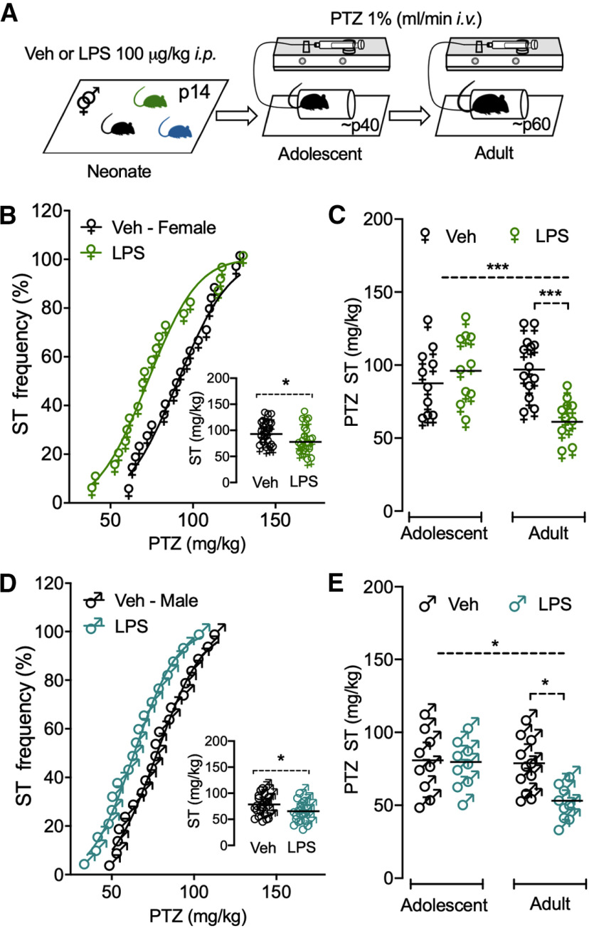
Early-life inflammatory stress increases seizure susceptibility later in life. However, possible sex- and age-specific differences and the associated mechanisms are largely unknown. C57BL/6 mice were bred in house, and female and male pups were injected with lipopolysaccharide (LPS; 100 μg/kg, i.p.) or vehicle control (saline solution) at postnatal day 14 (P14). Seizure threshold was assessed in response to pentylenetetrazol (1% solution, i.v.) in adolescence (∼P40) and adulthood (∼P60). We found that adult, but not adolescent, mice treated with LPS displayed ∼34% lower seizure threshold compared with controls. Females and males showed similar increased seizure susceptibility, suggesting that altered brain excitability was age dependent, but not sex dependent. Whole-cell recordings revealed no differences in excitatory synaptic activity onto CA1 pyramidal neurons from control or neonatally inflamed adolescent mice of either sex. However, adult mice of both sexes previously exposed to LPS displayed spontaneous EPSC frequency approximately twice that of controls, but amplitude was unchanged. Although these changes were not associated with alterations in dendritic spines or in the NMDA/AMPA receptor ratio, they were linked to an increased glutamate release probability from Schaffer collateral, but not temporoammonic pathway. This glutamate increase was associated with reduced activity of presynaptic GABAB receptors and was independent of the endocannabinoid-mediated suppression of excitation. Our new findings demonstrate that early-life inflammation leads to long-term increased hippocampal excitability in adult female and male mice associated with changes in glutamatergic synaptic transmission. These alterations may contribute to enhanced vulnerability of the brain to subsequent pathologic challenges such as epileptic seizures.SIGNIFICANCE STATEMENT Adult physiology has been shown to be affected by early-life inflammation. Our data reveal that early-life inflammation increases excitatory synaptic transmission onto hippocampal CA1 pyramidal neurons in an age-dependent manner through disrupted presynaptic GABAB receptor activity on Schaffer collaterals. This hyperexcitability was seen only in adult, and not in adolescent, animals of either sex. The data suggest a maturation process, independent of sex, in the priming action of early-life inflammation and highlight the importance of studying mature brains to reveal cellular changes associated with early-life interventions.

Keywords: Schaffer collaterals; age differences; glutamatergic transmission; hippocampus; lipopolysaccharide; presynaptic GABAB receptors.


Figures

Figure 1.
Figure 1.
Early-life inflammation reduced seizure threshold to the proconvulsive agent PTZ in adult, but not adolescent, female and male mice. A, Schematic showing experimental inflammatory paradigm; mice of both sexes were injected with LPS or Veh at P14, then returned to the home cage. Adolescent (∼P40) and adult (∼P60) mice received intravenous infusion of PTZ, which was terminated when a tonic–clonic seizure appeared. B, D, Cumulative probability of seizure threshold (ST) frequency in all female (B) and male (D) mice indicates a left ward shift (reduction) in a dose of PTZ required to elicit a seizure. Insets, Plots depicting amount of PTZ per kilogram of body weight necessary to induce a tonic–clonic seizure in all female (♀) and male (♂) mice. C, E, Summary data on the right show that increased seizure vulnerability (i.e., reduced PTZ dose) was in LPS-treated adult female (C) and male mice (E) compared with controls. No significant differences were observed in seizure threshold in adolescent mice exposed in early life to Veh or LPS. Each symbol represents one animal and the horizontal lines are the mean (♀ N = 44, ♂ N = 38). *p < 0.05, ***p < 0.001.
Figure 2.
Figure 2.
Early-life inflammation increased sEPSC frequency in adult, but not adolescent, mice. A, C, Representative traces of spontaneous synaptic activity of CA1 pyramidal cells from female (A) and male (C) mice previously injected with Veh (dark traces) or LPS (green/blue traces) at P14. Calibration: 20 pA; top trace for each color, 1 s; bottom trace, 0.1 s. B, D, Cumulative probability distribution of sEPSC frequency (left) and amplitude (right) of pyramidal neurons from female mice (B) and male mice (D). Insets show frequency and amplitude plotted for individual cells. Summary data display no significant differences in sEPSC amplitude and frequency in adolescent mice early exposed to Veh (white symbols) or LPS (green/blue symbols) at P14. Nevertheless, augmented sEPSC frequency, but not amplitude, was found in LPS-treated adult female and male mice compared with controls. Horizontal lines are the mean (n = 25–28 cells, 5–8 mice/group). SO, Stratum oriens; PTX, picrotoxin. *p < 0.05, ***p < 0.001.
Figure 3.
Figure 3.
Dendritic spine density of CA1 pyramidal neurons from adult mice is unaltered by early-life inflammation. A, B, Scatter graphs show no significant differences in spine density, number of bifurcated spines and number of spines with well defined heads of CA1 pyramidal cells from adult female (green symbols) and male (blue) mice early exposed to LPS compared with controls (white symbols). Each symbol represents one cell, and the horizontal lines are the mean (n = 13–19 cells, 3-4 mice/group).
Figure 4.
Figure 4.
Early-life inflammation selectively increased the probability of glutamate release in adult mice. A–D, Representative scaled traces of paired pulse recording of CA1 pyramidal cells evoked by the stimulation of afferent fibers in the Schaffer collateral (A, B) or temporoammonic (C, D) pathway (as indicated on the schematic on the left) from adult female (A, C) and male (B, D) mice exposed early in life to Veh (dark traces) or LPS (green/blue traces) at P14. Calibration: 100 pA, 50 ms. Scatter graphs on right show PPR of individual cells. Summary data display decreased PPR in CA1 pyramidal cells from both female (green symbols) and male (blue symbols) mice exposed to early inflammation compared with controls (white symbols) when the Schaffer collateral (A, B) was stimulated. However, when the temporoammonic pathway was stimulated (C, D), no significant differences in PPR were observed. Horizontal lines are the mean (n = 18–25 cells, 5–8 mice/group). SO, Stratum oriens; PTX, picrotoxin. **p < 0.01.
Figure 5.
Figure 5.
AMPA/NMDA ratio was not affected by early inflammation induced by LPS. A, B, Sample scaled traces of AMPA currents recorded at −70 mV and NMDA currents at +40 mV of CA1 pyramidal cells evoked by stimulation of afferent fibers in the Schaffer collateral pathway (as indicated on schematic on left) from adult female (A) and male (B) mice early exposed to Veh (dark traces) or LPS (green/blue traces) at P14. Calibration: 100 pA, 50 ms. Scatter graphs on right show the AMPA/NMDA ratio of individual cells. Summary data display no significant differences in the AMPA/NMDA ratio in pyramidal neurons from both female (green symbols) and male (blue symbols) mice early exposed to inflammation compared with controls (white symbols). Horizontal lines are the mean (n = 13–19 cells, 7–8 mice/group). SO, Stratum oriens; PTX, picrotoxin.
Figure 6.
Figure 6.
Early-life inflammation did not modify the DSE in adult CA1 pyramidal neurons. A, B, Representative traces of eEPSC recording (baseline: 1, immediately after 10 s postsynaptic depolarization: 2, and 1 min later: 3, corresponding to the indicated shaded region below) of CA1 pyramidal cells evoked by stimulation in the Schaffer collaterals from adult female (A) and male (B) mice exposed in early life to Veh (dark traces) or LPS (green/blue traces) at P14. Far right superimposed traces show responses in the presence and absence of the CB1 receptor antagonist AM251. Calibration: 200 pA, 100 ms. Bottom, Time course of eEPSC amplitude following postsynaptic depolarization to 0 mV for 5 or 10 s for Veh- or LPS-pretreated groups as well as after AM251 (2 µΜ) was added to block DSE (gray symbols). C, Summary data corresponding to shaded region in A and B showing that depression of eEPSC induced by 10 s depolarization is not significantly different between control and LPS pretreated CA1 pyramidal neurons from adult female (p = 0.96, top) and male mice (p = 0.99, bottom). n = 13–27 cells, 5–6 mice/group. ****p < 0.0001, ns, not significant.
Figure 7.
Figure 7.
Early-life inflammation diminished presynaptic GABAB receptor activity in adult mice. A, Top, Diagram showing synaptic cleft in presence of the GABAB receptor agonist baclofen in a vehicle-treated adult pyramidal cell; normal GABAB receptor activity maintains low presynaptic glutamate release and low postsynaptic spontaneous EPSC frequency (f). Bottom, Diagram illustrates that baclofen acting on GABAB receptors is less effective (dotted line) to decrease presynaptic glutamate release and postsynaptic spontaneous EPSC f in adult neonatally inflamed pyramidal cells. B, Representative scaled traces of paired pulse recording (with/without baclofen) of CA1 pyramidal cells evoked by stimulation of afferent fibers in the Schaffer collateral pathway from adult female (left) and male (right) mice exposed in early life to Veh (dark traces) or LPS (green/blue traces). Calibration: 200 pA, 20 ms. C, D, Scatter graphs on left show PPR of individual cells (with/without baclofen) and violin graphs on right show the comparison of the percentage of change among groups in females (C) and males (D). E, F, Pearson correlation coefficients between the baclofen-induced decrease in eEPSC amplitude and baclofen-induced increase in PPR from control and inflamed CA1 pyramidal neurons of female (E) and male mice (F). n = 10–11 cells, 4–5 mice/group. **p < 0.01, ***p < 0.001, ****p < 0.0001.
Figure 8.
Figure 8.
Early-life inflammation augmented spontaneous synaptic currents by decreasing presynaptic GABAB receptor activity in adult mice. A, B, Representative traces of spontaneous EPSCs (with/without baclofen) of CA1 pyramidal neurons from adult female (A) and male (B) mice previously injected with Veh (dark traces) or LPS (green/blue traces) at P14 (top trace and after addition of baclofen). Calibration: 20 pA, 200 ms. C–F, Scatter graphs on left show amplitude and frequency for individual cells (with/without baclofen) and violin graphs on right show comparison of the percentages of change among groups in females (C, E) and males (D, F). n = 10–11 cells, 4–5 mice/group. **p < 0.01, ***p < 0.001.

References

    1. Aboubakr A, Chait J, Lurie J, Schanzer HR, Marin ML, Faries PL, Ting W (2019) Secondary interventions after iliac vein stenting for chronic proximal venous outflow obstruction. J Vasc Surg Venous Lymphat Disord 7:670–676. 10.1016/j.jvsv.2019.02.016 - DOI - PubMed
    1. Acharjee S, Verbeek M, Gomez CD, Bisht K, Lee B, Benoit L, Sharkey KA, Benediktsson A, Tremblay ME, Pittman QJ (2018) Reduced microglial activity and enhanced glutamate transmission in the basolateral amygdala in early CNS autoimmunity. J Neurosci 38:9019–9033. 10.1523/JNEUROSCI.0398-18.2018 - DOI - PMC - PubMed
    1. Aksoy-Aksel A, Manahan-Vaughan D (2013) The temporoammonic input to the hippocampal CA1 region displays distinctly different synaptic plasticity compared to the Schaffer collateral input in vivo: significance for synaptic information processing. Front Synaptic Neurosci 5:5. 10.3389/fnsyn.2013.00005 - DOI - PMC - PubMed
    1. Auvin S, Shin D, Mazarati A, Sankar R (2010) Inflammation induced by LPS enhances epileptogenesis in immature rat and may be partially reversed by IL1RA. Epilepsia 51:34–38. 10.1111/j.1528-1167.2010.02606.x - DOI - PMC - PubMed
    1. Banks WA, Gray AM, Erickson MA, Salameh TS, Damodarasamy M, Sheibani N, Meabon JS, Wing EE, Morofuji Y, Cook DG, Reed MJ (2015) Lipopolysaccharide-induced blood-brain barrier disruption: roles of cyclooxygenase, oxidative stress, neuroinflammation, and elements of the neurovascular unit. J Neuroinflammation 12:223. 10.1186/s12974-015-0434-1 - DOI - PMC - PubMed

Publication types