. 2021 Jun 10;95(13):e0197420.
doi: 10.1128/JVI.01974-20. Epub 2021 Jun 10.

ApoA1 Neutralizes Proinflammatory Effects of Dengue Virus NS1 Protein and Modulates Viral Immune Evasion

Affiliations

ApoA1 Neutralizes Proinflammatory Effects of Dengue Virus NS1 Protein and Modulates Viral Immune Evasion

Diego R Coelho et al. J Virol. .

Abstract

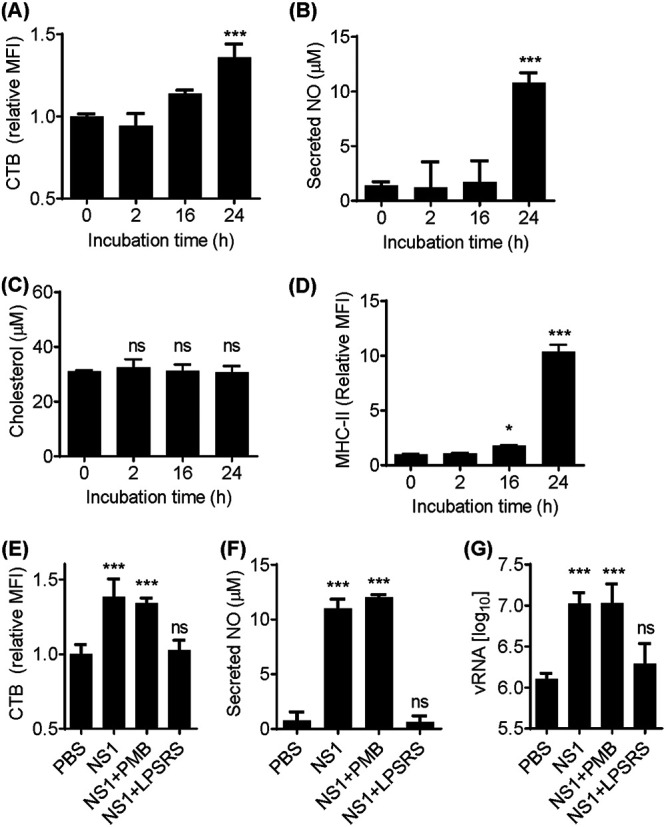
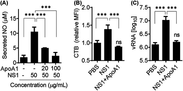
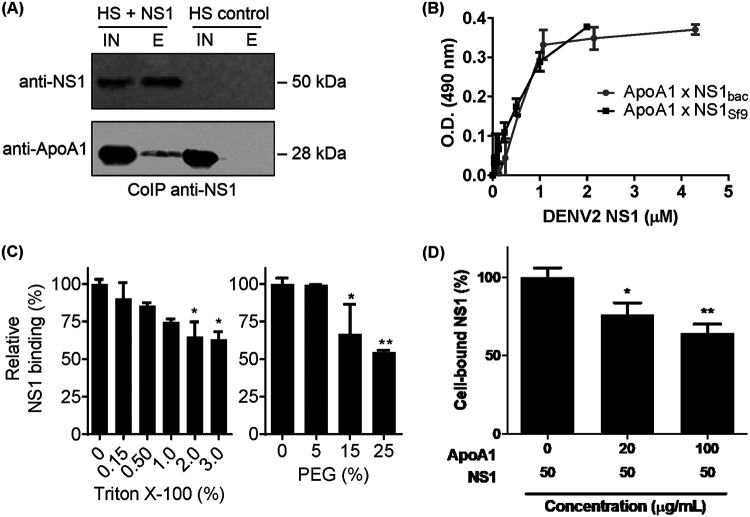
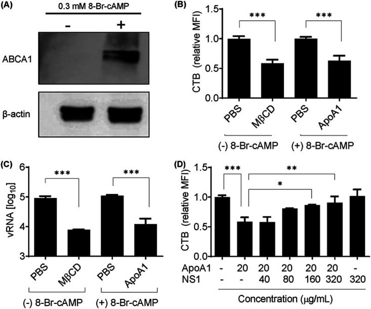
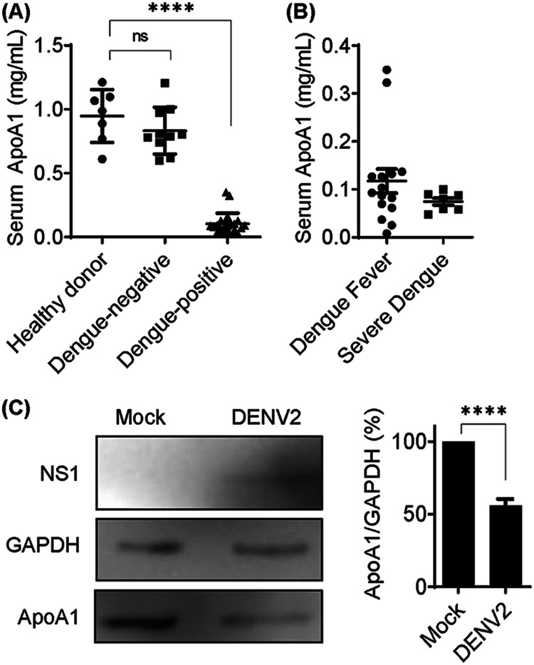
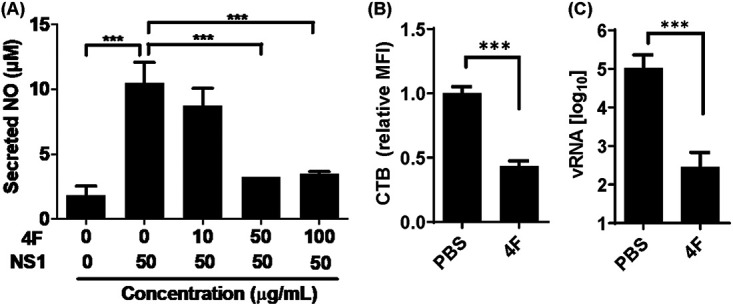
Dengue is a mosquito-borne infectious disease that is highly endemic in tropical and subtropical countries. Symptomatic patients can rapidly progress to severe conditions of hemorrhage, plasma extravasation, and hypovolemic shock, which leads to death. The blood tests of patients with severe dengue typically reveal low levels of high-density lipoprotein (HDL), which is responsible for reverse cholesterol transport (RCT) and regulation of the lipid composition in peripheral tissues. It is well known that dengue virus (DENV) depends on membrane cholesterol rafts to infect and to replicate in mammalian cells. Here, we describe the interaction of DENV nonstructural protein 1 (NS1) with apolipoprotein A1 (ApoA1), which is the major protein component of HDL. NS1 is secreted by infected cells and can be found circulating in the serum of patients with the onset of symptoms. NS1 concentrations in plasma are related to dengue severity, which is attributed to immune evasion and an acute inflammatory response. Our data show that the DENV NS1 protein induces an increase of lipid rafts in noninfected cell membranes and enhances further DENV infection. We also show that ApoA1-mediated lipid raft depletion inhibits DENV attachment to the cell surface. In addition, ApoA1 is able to neutralize NS1-induced cell activation and to prevent NS1-mediated enhancement of DENV infection. Furthermore, we demonstrate that the ApoA1 mimetic peptide 4F is also capable of mediating lipid raft depletion to control DENV infection. Taken together, our results suggest the potential of RCT-based therapies for dengue treatment. These results should motivate studies to assess the importance of RCT in DENV infection in vivo. IMPORTANCE DENV is one of the most relevant mosquito-transmitted viruses worldwide, infecting more than 390 million people every year and leading to more than 20 thousand deaths. Although a DENV vaccine has already been approved, its potential side effects have hampered its use in large-scale immunizations. Therefore, new treatment options are urgently needed to prevent disease worsening or to improve current clinical management of severe cases. In this study, we describe a new interaction of the NS1 protein, one of the major viral components, with a key component of HDL, ApoA1. This interaction seems to alter membrane susceptibility to virus infection and modulates the mechanisms triggered by DENV to evade the immune response. We also propose the use of a mimetic peptide named 4F, which was originally developed for atherosclerosis, as a potential therapy for relieving DENV symptoms.

Keywords: apolipoprotein A1; dengue virus; immune evasion; nonstructural 1 protein; protein-protein interactions.


Figures

FIG 1
FIG 1
NS1-induced cell activation promotes lipid raft accumulation on the cell membrane and facilitates DENV attachment. (A) RAW 264.7 cells were incubated with 50 μg/ml NS1 for up to 24 h. Lipid rafts were quantified by flow cytometry using CTB-FITC, and the mean fluorescence intensity (MFI) was calculated relative to control cells incubated with PBS. (B) Immune activation was assessed by quantification of secreted NO in culture supernatants. (C) Total cholesterol was quantified in cell extracts from NS1-treated cells. (D) MHC-II expression on the cell surface was assessed by flow cytometry of NS1-treated cells. (E) Cells were treated with NS1 for 24 h in the presence of the LPS inhibitor PMB (100 μg/ml) or the LPS competitor LPS-RS (100 μg/ml). Lipid rafts were assessed by CTB -FITC incorporation. (F) Secreted NO was quantified in the supernatants by the Griess assay. (G) NS1-treated cells were challenged with DENV2 for 1 h at 4°C for viral attachment. Unbound virus was washed out with PBS, and total RNA was extracted. vRNA was quantified by qRT-PCR. Error bars represent the SDs of at least three biological replicates. Asterisks represent significant differences, compared to control. *, P < 0.1; ***, P < 0.001; ns, not significant.
FIG 2
FIG 2
ApoA1 inhibits NS1-induced cell activation and the NS1 facilitation effect on DENV2 attachment to the cell membrane. (A) RAW 264.7 cells were incubated with NS1 for 24 h in the presence of different concentrations of ApoA1. Secreted NO was quantified in culture supernatants by the Griess assay. (B) Cells were fixed and labeled with CTB-FITC, and lipid rafts were analyzed by flow cytometry. The MFI of CTB was quantified relative to control cells incubated with PBS (pH 7.4). (C) Cells were preincubated for 24 h with 50 μg/ml NS1, in the presence or absence of 100 μg/ml ApoA1, and then challenged with DENV2 for 1 h at 4°C for virus attachment. Unbound virus was washed out with PBS, and total RNA was extracted. vRNA was quantified by qRT-PCR. Error bars represent the SDs of at least three biological replicates. Asterisks represent significant differences, compared to control. ***, P < 0.001; ns, not significant.
FIG 3
FIG 3
NS1 interacts with ApoA1 via nonpolar interfaces. (A) Purified NS1 was incubated with 10% human serum (HS) and immunoprecipitated with purified anti-NS1 antibody. The input (IN) and elution (E) fractions were analyzed by Western blotting with anti-NS1 and anti-ApoA1 monoclonal antibodies. (B) Microplates were coated with purified ApoA1 and incubated with increasing amounts of recombinant NS1 protein expressed in bacteria (NS1bac) or in Sf9 cells (NS1Sf9). Bound NS1 was detected using anti-NS1 polyclonal antibody and HRP-conjugated secondary antibody. OD490 values were normalized to negative-control (BSA) values. (C) ApoA1-coated microplates were incubated with 0.6 μM NS1bac in the presence of Triton X-100 or PEG. Bound NS1 was detected as described previously. (D) A monolayer of RAW 264.7 cells was fixed with 4% paraformaldehyde, blocked, and coincubated with purified ApoA1 and NS1Sf9. Bound NS1 was detected using anti-NS1 polyclonal antibody and HRP-conjugated secondary antibody. OD490 values were normalized to negative-control (BSA) values. Error bars indicate SDs of two independent experiments. *, P < 0.5; **, P < 0.05.
FIG 4
FIG 4
ApoA1-mediated lipid raft depletion from ABCA1-expressing cells inhibits DENV2 attachment to the cell membrane. (A) RAW 264.7 cells were treated with 0.3 mM 8-Br-cAMP for 16 h. Total cell lysate was analyzed by Western blotting with anti-ABCA1. (B) Lipid rafts were quantified by flow cytometry of cells labeled with CTB-FITC. Control cells were treated with PBS (pH 7.4) or depleted with 3 mM MβCD for 2 h. ABCA1-expressing cells were treated with 100 μg/ml ApoA1 for 2 h. MFI was calculated relative to control cells. (C) After induced lipid raft depletion, RAW 264.7 cells were challenged with DENV2 for 1 h at 4°C for viral attachment. Unbound virus was washed out with PBS, and total RNA was extracted. vRNA was quantified by qRT-PCR. (D) ABCA1-expressing cells were incubated with ApoA1 for 2 h in the presence of different concentrations of NS1. Lipid rafts were quantified by flow cytometry of CTB-FITC. Each bar represents the SD of at least three biological replicates. Asterisks represent significant differences, compared to control. *, P < 0.01; **, P < 0.01; ***, P < 0.001.
FIG 5
FIG 5
Serum ApoA1 concentrations are decreased in DENV-infected patients. (A) Total ApoA1 was quantified in human serum using the human ApoA1 ELISA kit (Thermo Fisher Scientific). Of samples from 33 patients, 23 were DENV positive and 10 were DENV negative. Samples from 7 healthy donors were also quantified as control samples. (B) Among DENV-positive patients, 16 were diagnosed with dengue fever and 7 were diagnosed with severe dengue. Bars represent mean and SDs. (C) HepG2 cells were mock infected or infected with DENV2 (multiplicity of infection of 1) for 48 h, and total cell lysates were analyzed by Western blotting. The ApoA1 band was quantified and expressed relative to glyceraldehyde-3-phosphate dehydrogenase (GAPDH). Bars represent means and SDs from three experiments. Asterisks represent significant differences, compared to mock-infected cells. ****, P < 0.005; ns, not significant.
FIG 6
FIG 6
Peptide 4F-mediated lipid raft depletion from ABCA1-expressing cells inhibits DENV attachment to the cell membrane. (A) RAW 264.7 cells were incubated with NS1 for 24 h, in the presence of different concentrations of peptide 4F. Secreted NO was quantified from culture supernatants by the Griess assay. (B) ABCA1-expressing cells were incubated with 50 μg/ml peptide 4F for 2 h. Cells were fixed and labeled with CTB-FITC, and lipid rafts were analyzed by flow cytometry. The MFI of CTB was quantified relative to control cells incubated with PBS (pH 7.4). (C) ABCA1-expressing cells were incubated with peptide 4F for 2 h and then challenged with DENV2 for 1 h at 4°C for viral attachment. Unbound virus was washed out with PBS, and total RNA was extracted. vRNA was quantified by qRT-PCR. Error bars represent the SDs of at least three independent experiments. Asterisks represent significant differences, compared to control. ***, P < 0.001.

References

    1. Guzman MG, Harris E. 2015. Dengue. Lancet 385:453–465. doi:10.1016/S0140-6736(14)60572-9. - DOI - PubMed
    1. Flipse J, Smit JM. 2015. The complexity of a dengue vaccine: a review of the human antibody response. PLoS Negl Trop Dis 9:e0003749. doi:10.1371/journal.pntd.0003749. - DOI - PMC - PubMed
    1. Swaminathan S, Khanna N. 2019. Dengue vaccine development: global and Indian scenarios. Int J Infect Dis 84:S80–S86. doi:10.1016/j.ijid.2019.01.029. - DOI - PubMed
    1. World Health Organization. 2009. Dengue guidelines for diagnosis, treatment, prevention and control. World Health Organization, Geneva, Switzerland. - PubMed
    1. Chaloemwong J, Tantiworawit A, Rattanathammethee T, Hantrakool S, Chai-Adisaksopha C, Rattarittamrong E, Norasetthada L. 2018. Useful clinical features and hematological parameters for the diagnosis of dengue infection in patients with acute febrile illness: a retrospective study. BMC Hematol 18:20. doi:10.1186/s12878-018-0116-1. - DOI - PMC - PubMed

Publication types