. 2021 Apr 7;12(4):374.
doi: 10.1038/s41419-021-03657-0.

Mutation-specific non-canonical pathway of PTEN as a distinct therapeutic target for glioblastoma

Affiliations

Mutation-specific non-canonical pathway of PTEN as a distinct therapeutic target for glioblastoma

Seung Won Choi et al. Cell Death Dis. .

Abstract

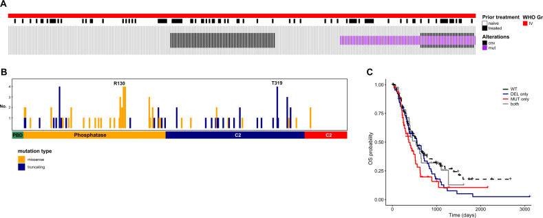
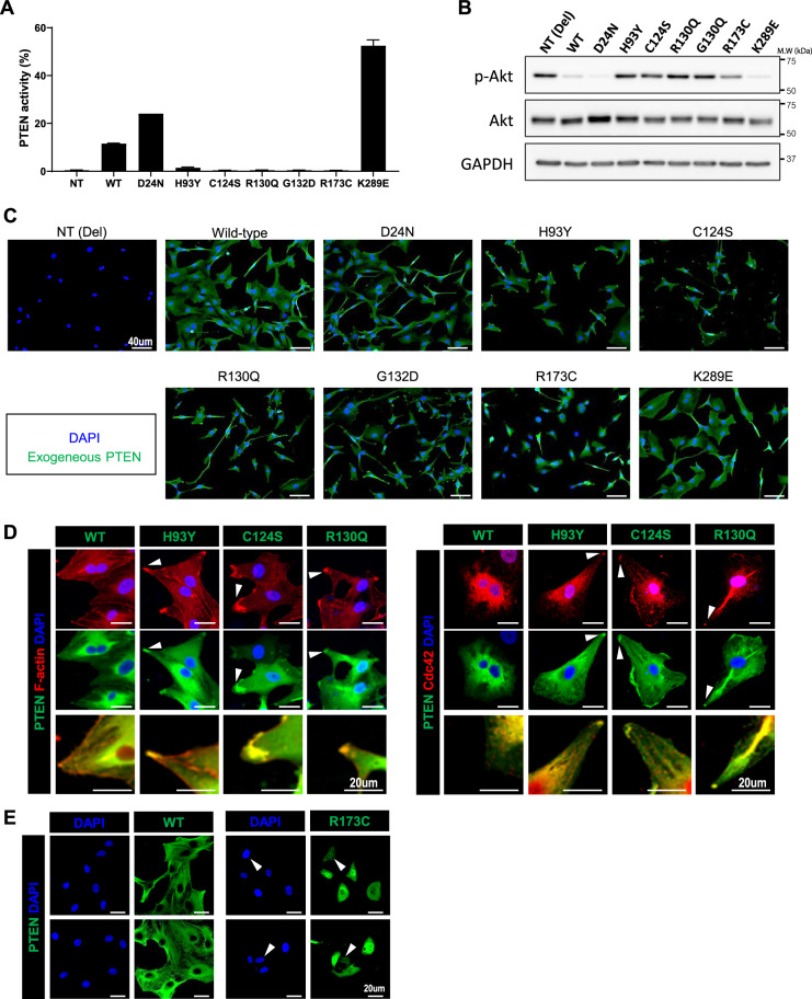
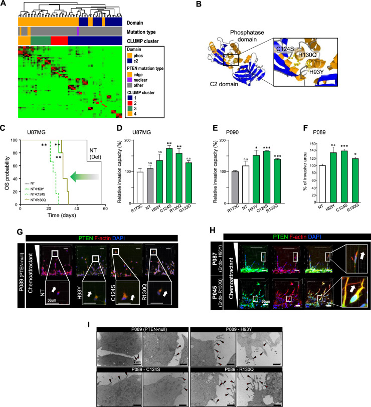
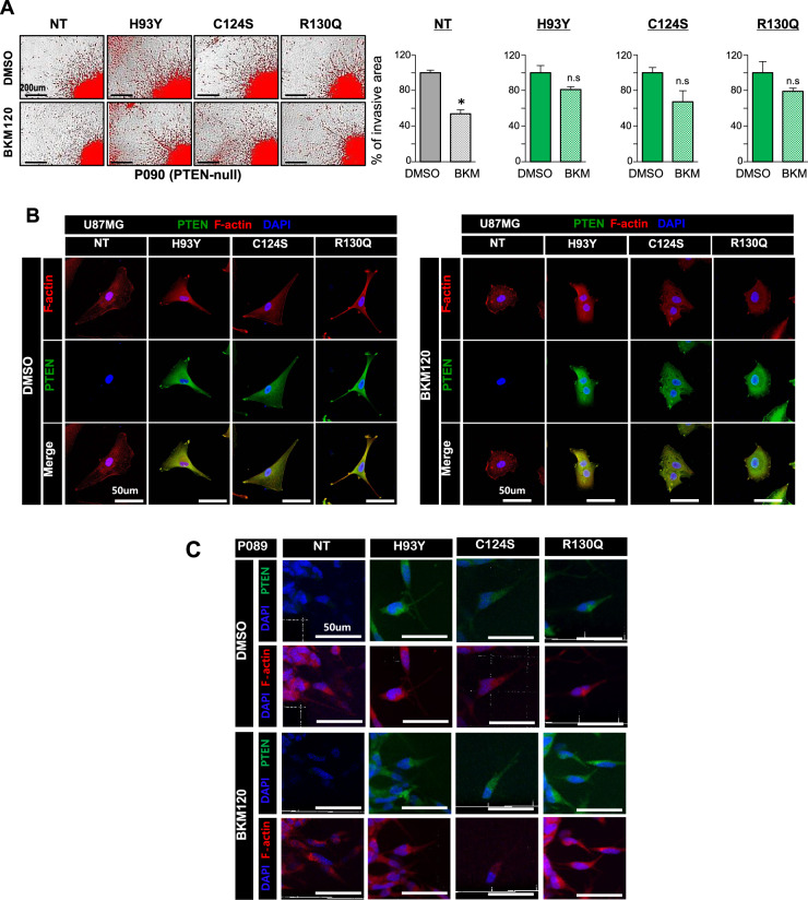
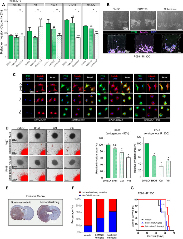
PTEN is one of the most frequently altered tumor suppressor genes in malignant tumors. The dominant-negative effect of PTEN alteration suggests that the aberrant function of PTEN mutation might be more disastrous than deletion, the most frequent genomic event in glioblastoma (GBM). This study aimed to understand the functional properties of various PTEN missense mutations and to investigate their clinical relevance. The genomic landscape of PTEN alteration was analyzed using the Samsung Medical Center GBM cohort and validated via The Cancer Genome Atlas dataset. Several hotspot mutations were identified, and their subcellular distributions and phenotypes were evaluated. We established a library of cancer cell lines that overexpress these mutant proteins using the U87MG and patient-derived cell models lacking functional PTEN. PTEN mutations were categorized into two major subsets: missense mutations in the phosphatase domain and truncal mutations in the C2 domain. We determined the subcellular compartmentalization of four mutant proteins (H93Y, C124S, R130Q, and R173C) from the former group and found that they had distinct localizations; those associated with invasive phenotypes ('edge mutations') localized to the cell periphery, while the R173C mutant localized to the nucleus. Invasive phenotypes derived from edge substitutions were unaffected by an anti-PI3K/Akt agent but were disrupted by microtubule inhibitors. PTEN mutations exhibit distinct functional properties regarding their subcellular localization. Further, some missense mutations ('edge mutations') in the phosphatase domain caused enhanced invasiveness associated with dysfunctional cytoskeletal assembly, thus suggesting it to be a potent therapeutic target.


Conflict of interest statement

The authors declare no competing interests.

Figures

Fig. 1
Fig. 1. Genomic landscape of PTEN in glioblastoma patients.
A Genomic landscape of PTEN in glioblastoma (GBM) in the study cohort. B Mutation profiles of PTEN alterations in GBM tumors. Statistically significant association between mutation type and PTEN domain was identified; missense mutations were dominant in the phosphatase domain, while truncating mutations were prevalent in the C2 domain (p-value < 0.001, Fisher’s exact test). C Survival outcome of GBM patients stratified by PTEN alteration status. There was no statistically significant difference in prognosis as a function of the alteration status of PTEN (p-value = 0.1, Log-rank test). Survival analysis was performed using Kaplan–Meier method and log-rank test was used to statistically compare the curves. cnv, copy-number variation; mut, mutation; no., number; OS, overall survival; WT, wild-type; DEL-only, deletion only; MUT-only, mutation only; both, both mutation and deletion alleles.
Fig. 2
Fig. 2. Distinct subcellular compartmentalization of PTEN mutants.
We established cell lines overexpressing different PTEN mutants of interest using U87MG cells. A PTEN activity was measured by converted ratio of PI-,-P2 from supplemented PI--P3 by PTEN protein in U87MG cell lines with exogenous PTEN mutants. Data are shown as the means of triplicates of experiments ± s.d. B p-Akt activity of U87MG cell lines expressing wild-type PTEN and PTEN mutants. C Subcellular compartmentalization differed by PTEN mutations—some mutations (H93Y, C124S, and R130Q) with cytoplasmic distribution exhibited peculiar localization at cell periphery, while another mutation (R173C) showed nuclear localization. D PTEN mutants located in the cytoplasmic compartment co-localized with markers of the leading edge in migrating cells, Cdc42 and F-actin (white arrow). High-resolution images depicting the co-staining of PTEN and F-actin are presented at the bottommost row. E Nuclear compartmentalization of the R173C PTEN mutant (white arrow). WT, wild-type; NT, null-type; Del, deletion; s.d: standard deviation.
Fig. 3
Fig. 3. Invasive phenotype of PTEN edge mutants.
U87MG cells and PTEN-null PDCs (P089 and P090) were used to overexpress PTEN edge mutants. PDCs with endogenous PTEN edge mutation were also used for validation (P045 with R130Q mutation; P087 with H93Y mutation). A CLUMP analysis of PTEN mutations identified in SMC and TCGA cohorts. A total of 68 residues were finally selected for CLUMP analysis and CLUMP analysis revealed four distinct subgroups of PTEN mutations. According to CLUMP analysis, PTEN mutations within the phosphatase domain were further divided into distinct subgroups, which correlated with the 3D structure of PTEN protein. B 3D crystal structure of the PTEN protein: all residues changed by PTEN edge mutations (H93, C124, and R130) are located within the same pocket of the phosphatase domain. C Comparison of survival outcomes of orthotopic xenografts established by U87MG cells with distinct PTEN mutants. The mice with edge mutations showed poor survival outcome compared to PTEN-null mice (P-value <0.001, Log-rank test). Three PTEN-null xenografts were used as control group, while six PTEN-mutant xenografts were made per each mutation of interest. DF In vitro invasion assay to assess the enhanced invasion capacity of PTEN edge mutation compared to nuclear mutation. Data are shown as the means of triplicates of experiments ± s.d. The P-value was calculated by two-sided t test. D In vitro trans-well invasion assay using U87MG cells with exogenous mutants. All tumor cells with edge mutants exhibited significantly increased invasion capacity compared to tumors with nuclear mutants, except H93Y (p-value=0.076, 0.005, and 0.008 for H93Y, C124S, and R130Q, respectively, two-sided t test). Their enhanced invasion capacity was also significantly superior to PTEN-null U87MG cells (p-value=0.203, 0.001, and 0.015 for H93Y, C124S, and R130Q, respectively, two-sided t test). E in vitro 3D sphenoid invasion assay using PTEN-null PDCs (P090) with exogenous mutants. All PDCs (P090) with edge mutants exhibited significantly enhanced invasion capacity compared to PDCs with nuclear mutation (p-value=0.028, <0.001, and <0.001 for H93Y, C124S, and R130Q, respectively, two-sided t test). F In vitro microfluid assay to measure the development of invasion in response to chemotactic stimuli in PTEN-null PDCs (P089) with exogenous mutants. PDCs expressing edge mutants showed a more invasive phenotype than PTEN-null PDCs (p-value=0.135, <0.001, and 0.023 for H93Y, C124S, and R130Q, respectively, two-sided t test). G, H Co-localization of F-actin and PTEN edge mutants at the cell periphery of motile PDCs upon chemotactic stimuli. I Development of cellular projections (black arrow) of PDCs (P089) with exogenous PTEN edge mutations. NT, null-type; Del, deletion.
Fig. 4
Fig. 4. Invasive phenotypes of PTEN edge mutants are not altered by BKM120, a PI3K/Akt inhibitor.
A Development of invasive branches in PTEN-null PDCs (P090) overexpressing PTEN edge mutation was not altered by BKM120, a pan-PI3K inhibitor (p-value = <0.001, 0.091, 0.071, and 0.193 for NT, H93Y, C124S, and R130Q, two-sided t test). Data are shown as the means of four experimental replicates ± s.d. B, C Persistence of PTEN edge mutants at the cellular edge of U87MG cells and PTEN-null PDCs (P089) with exogenous edge mutation following BKM120 treatment. NT, null-type; del, deletion; conc., concentration.
Fig. 5
Fig. 5. A microtubule inhibitor disrupts invasive phenotypes of PTEN edge mutants in vitro and in vivo.
A Decrease in invasion capacity following microtubule inhibitor treatment (Colchicine) in PTEN-null PDCs (P090) overexpressing PTEN edge mutants. Data are shown as the means of triplicates of experiments ± s.d. PDCs expressing edge mutants exhibited significantly fewer invasive cellular projections following colchicine treatment (p-value=0.009, <0.001, and <0.001 for H93Y, C124S, and R130Q, one-sided t test). B Decrease in invasion following microtubule inhibitor treatment in PTEN-null PDCs with exogenous edge mutation (P089-R130Q) observed in microfluid assay; morphological changes captured during therapy are presented below, which reflecting less invasive property. C Development of tubulin aggresomes after microtubule inhibitor treatment in U87MG cells overexpressing PTEN edge mutations. D Decreased invasive branches of PDCs with endogenous edge mutation following microtubule inhibitor treatment. Data are shown as the means of four experimental replicates ± s.d. PDCs with endogenous edge mutations exhibited significantly decreased invasion following colchicine treatment than BKM120 (p-value=0.114, 0.029, and 0.029 for BKM120, Col, and Vin, respectively (P087); p-value=0.029, 0.029, and 0.029 for BKM120, Col, and Vin, respectively (P045), two-sided Wilcoxon test). EG Intracranial xenograft model was established using PTEN-null PDCs with exogenous edge mutants (P090-R130Q); Five xenografts were treated by vehicle, while seven and eight mice were treated by BKM120 and Colchicine, respectively. E Invasive scoring system to evaluate the severity of invasion in vivo. We redefined the original invasion scoring system into binary classification, “non-invasive/mild” versus “moderate/strong”. The extent of tumor cell transmission via corpus callosum is the main criteria to distinguish the severity of invasion. F Reduction in infiltrative extent following microtubule inhibitor treatment in in vivo models. Colchicine-treated group exhibited greater decrease in invasiveness than BKM120-treated group, however, this was not statistically significant (p-value=0.39, Fisher’s exact test). G Orthotopic xenografts treated by microtubule inhibitor (colchicine) exhibited improved survival outcome compared to vehicle-treated xenografts, while BKM120 treatment failed to show statistically significant improvement of prognosis. Survival analysis was performed using a Kaplan–Meier plot and the log-rank test was used to show statistical differences between survival curves. Col, colchicine; Vin, vinorelbine; n.s., non-significant.
Fig. 6
Fig. 6. Radiographic failure patterns of GBM tumors with PTEN edge mutations.
A Representative MR images depicting the locally invasive failure pattern of GBM tumors with PTEN edge mutation. Two GBM patients with PTEN edge mutation R130Q demonstrated locally invasive failure patterns, with tumors transmitted to the contralateral hemisphere in a locally invasive, infiltrative manner. B Distribution of radiographic recurrence patterns in CLUM-defined clusters. Edge mutations were included in cluster 4, while, the nuclear mutation was included in cluster 1 with other mutations in the C2 domain. LMC, leptomeningeal seeding.

Similar articles

Cited by

References

    1. Cancer Genome Atlas Research Network. Comprehensive genomic characterization defines human glioblastoma genes and core pathways. Nature. 2008;455:1061–1068. doi: 10.1038/nature07385. - DOI - PMC - PubMed
    1. Papa A, et al. Cancer-associated PTEN mutants act in a dominant-negative manner to suppress PTEN protein function. Cell. 2014;157:595–610. doi: 10.1016/j.cell.2014.03.027. - DOI - PMC - PubMed
    1. Liaw D, et al. Germline mutations of the PTEN gene in Cowden disease, an inherited breast and thyroid cancer syndrome. Nat. Genet. 1997;16:64–67. doi: 10.1038/ng0597-64. - DOI - PubMed
    1. Steck PA, et al. Identification of a candidate tumour suppressor gene, MMAC1, at chromosome 10q23.3 that is mutated in multiple advanced cancers. Nat. Genet. 1997;15:356–362. doi: 10.1038/ng0497-356. - DOI - PubMed
    1. Wang X, Jiang X. PTEN: a default gate-keeping tumor suppressor with a versatile tail. Cell Res. 2008;18:807–816. doi: 10.1038/cr.2008.83. - DOI - PubMed

Publication types