. 2021 Apr 7;11(1):7572.
doi: 10.1038/s41598-021-86746-6.

New insights into microstructure of neutron-irradiated tungsten

Affiliations

New insights into microstructure of neutron-irradiated tungsten

M Dürrschnabel et al. Sci Rep. .

Abstract

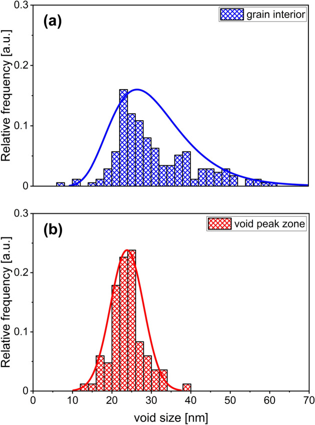
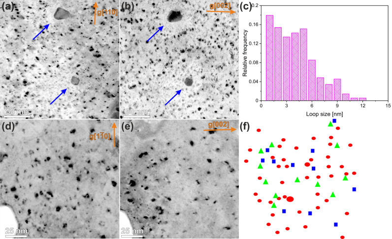
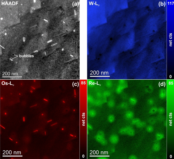
The development of appropriate materials for fusion reactors that can sustain high neutron fluence at elevated temperatures remains a great challenge. Tungsten is one of the promising candidate materials for plasma-facing components of future fusion reactors, due to several favorable properties as for example a high melting point, a high sputtering resistivity, and a low coefficient of thermal expansion. The microstructural details of a tungsten sample with a 1.25 dpa (displacements per atom) damage dose after neutron irradiation at 800 °C were examined by transmission electron microscopy. Three types of radiation-induced defects were observed, analyzed and characterized: (1) voids with sizes ranging from 10 to 65 nm, (2) dislocation loops with a size of up to 10 nm and (3) W-Re-Os containing σ- and χ-type precipitates. The distribution of voids as well as the nature of the occurring dislocation loops were studied in detail. In addition, nano-chemical analyses revealed that the σ- and χ-type precipitates, which are sometimes attached to voids, are surrounded by a solid solution cloud enriched with Re. For the first time the crystallographic orientation relationship of the σ- and χ-phases to the W-matrix was specified. Furthermore, electron energy-loss spectroscopy could not unambiguously verify the presence of He within individual voids.


Conflict of interest statement

The authors declare no competing interests.

Figures

Figure 1
Figure 1
Bright-field TEM images acquired from neutron-irradiated W. (a) A representative area of a grain interior with voids (bright spots) and Re–Os-rich precipitates (dark strips). The inset shows a magnified view of a individual void attached to a W–Re–Os-rich precipitate. (b) A representative GB region. The numbered areas are (1) the void denuded zone, (2) void-peak zone and (3) the second denuded zone.
Figure 2
Figure 2
Size distribution histograms of the voids registered in the (a) grain interior and (b) void peak zone.
Figure 3
Figure 3
Visualisation of dislocation loops and black dots using reverse contrast dark field (DF) imaging. DF images were obtained with g[110] (a, d) and g[002] (b, e) g-vectors near the [110] zone-axis. The size distribution histogram of the dislocation loops is shown in (c). The loops with b½〈111〉 and b〈100〉 Burgers vectors are imaged in the part (f) with red circles and blue squares, respectively. The loops with undefined Burgers vector are denoted by green triangles.
Figure 4
Figure 4
STEM-EDX spectrum image of a representative region in a neutron-irradiated W specimen. (a) STEM-HAADF image. Elemental maps of (b) W, (c) Os, and (d) Re display the location of elements with respect to the sample morphology. The tile-like structure in the HAADF image and the W map is surface topography due to flash polishing.
Figure 5
Figure 5
Results of a blind source separation (BSS) by PCA and ICA of the STEM-EDX data presented in Fig. 6. (a) Screen plot of the first 30 principal components (the dashed line indicates the number of independent components). The inset shows a schematic cross-sectional view of the sample for illustration purposes. (b) Corresponding independent component spectra. (c) Independent component maps showing the presence of Os-rich cores (IC#0), a W matrix (IC#1), a Re-rich cloud around the Os-rich cores (IC#2), and an oxide surface layer (IC#3), which is most probably due to the sample preparation.
Figure 6
Figure 6
STEM-EDX mapping near GB. (a) STEM-HAADF image with marked position of GB. Elemental maps of (b) W, (c) Os, and (d) Re display the location of elements with respect to the sample morphology. The profiles in parts (e) and (f) indicate the distribution of the Os and Re concentration near the GB.
Figure 7
Figure 7
(a) High-resolution phase contrast image of a spherical-shaped Os-rich precipitate, which is identified as the WOs2 σ-phase. Magnified view (b) and diffractogram (FFT) (c) of the region delimited by the orange square in (a). (d) Simulated model of the diffractogram.
Figure 8
Figure 8
(a) High-resolution phase contrast image of a rod-shaped Os-rich precipitate, which is identified as the WOs3 χ-Phase. Magnified view (b) and diffractogram (FFT) (c) of the region delimited by the orange square in (a). Simulated model of the diffractogram (d).
Figure 9
Figure 9
(a) TEM bright-field image of a faceted void attached to a W–Os rod. (b) High-resolution phase contrast image of the W–Os phase exhibiting Moiré fringes in the Os-rich part. (c) Diffractogram (FFT) of (b). (d) Composite STEM-EDX elemental map of the same sample region.
Figure 10
Figure 10
Combined STEM-EDX and low-loss STEM-EELS measurement of a individual void. The intensities of the STEM-EELS maps was obtained by NLLS fitting of the peaks located in the W plasmon peak area.

References

    1. Bolt H, et al. Materials for the plasma-facing components of fusion reactors. J. Nucl. Mater. 2004;329–333:66–73. doi: 10.1016/j.jnucmat.2004.04.005. - DOI
    1. Rieth M, Doerner R, Hasegawa A, Ueda Y, Wirtz M. Behavior of tungsten under irradiation and plasma interaction. J. Nucl. Mater. 2019;519:334–368. doi: 10.1016/j.jnucmat.2019.03.035. - DOI
    1. Lloyd MJ, et al. Decoration of voids with rhenium and osmium transmutation products in neutron irradiated single crystal tungsten. Scripta Mater. 2019;173:96–100. doi: 10.1016/j.scriptamat.2019.07.036. - DOI
    1. Hasegawa A, Fukuda M, Yabuuchi K, Nogami S. Neutron irradiation effects on the microstructural development of tungsten and tungsten alloys. J. Nucl. Mater. 2016;471:175–183. doi: 10.1016/j.jnucmat.2015.10.047. - DOI
    1. Hwang T, et al. Effect of neutron irradiation on rhenium cluster formation in tungsten and tungsten-rhenium alloys. J. Nucl. Mater. 2018;507:78–86. doi: 10.1016/j.jnucmat.2018.04.031. - DOI

LinkOut - more resources