

Characteristics of fixation patterns and their relationship with visual function of patients with idiopathic macular holes after vitrectomy
- PMID: 33828327
- PMCID: PMC8027455
- DOI: 10.1038/s41598-021-87286-9
Characteristics of fixation patterns and their relationship with visual function of patients with idiopathic macular holes after vitrectomy
Abstract
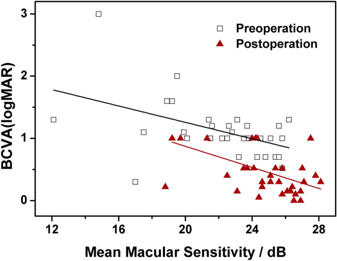
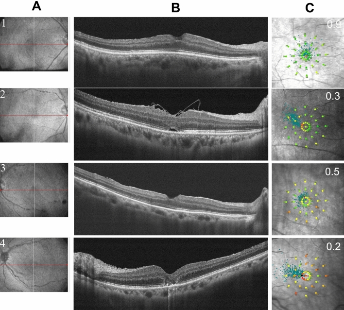
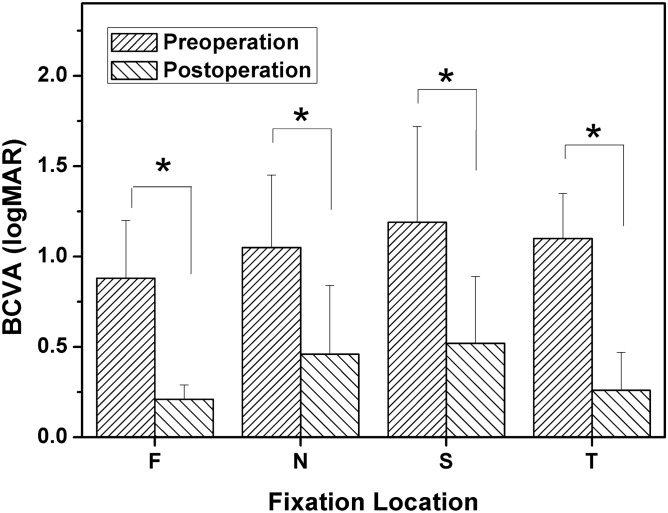
To analyze the relationships between the fixation location and the visual function of idiopathic macular hole (IMH) patients with macular integrity assessment (MAIA) examination preoperatively and 3 months postoperatively. This was a retrospective case analysis. Forty-three eyes of 43 patients diagnosed with IMH were included in this study. The best corrected visual acuity (BCVA) assessments, optical coherence tomography (OCT) and MAIA examinations were performed before surgery and 1 week, 1 month and 3 months after surgery. The relationships between MAIA parameters and visual acuity were assessed by correlation analysis. Grouping by fixation location with the foveola (2°) as the centre, the locations could be divided into five groups, including foveolar, temporal, nasal, inferior and superior fixation. The mean macular sensitivity (MMS) of the macular area was correlated with the BCVA in the IMH patients before and 3 months after surgery (before surgery P = 0.00, after surgery P = 0.00). The MMS could be used as a good indicator for evaluating visual function in IMH patients. There was a significant difference in fixation location before and after the operation (P = 0.01). The preoperative fixation location of IMH patients was mainly in the superior area, while postoperatively moved to the foveola and nasal areas. Paying attention to the changes of fixation locations in IMH patients may provide new clues for further improving postoperative visual function.
Conflict of interest statement
The authors declare no competing interests.
Figures

Similar articles
-
Effect of preoperative retinal sensitivity and fixation on long-term prognosis for idiopathic macular holes.Graefes Arch Clin Exp Ophthalmol. 2012 Nov;250(11):1587-96. doi: 10.1007/s00417-012-1997-5. Epub 2012 Mar 24. Graefes Arch Clin Exp Ophthalmol. 2012. PMID: 22441811
-
Morphologic and functional evaluation before and after successful macular hole surgery using spectral-domain optical coherence tomography combined with microperimetry.Retina. 2012 Oct;32(9):1733-42. doi: 10.1097/IAE.0b013e318242b81a. Retina. 2012. PMID: 22466479
-
OCT angiography quantifying choriocapillary circulation in idiopathic macular hole before and after surgery.Graefes Arch Clin Exp Ophthalmol. 2017 May;255(5):893-902. doi: 10.1007/s00417-017-3586-0. Epub 2017 Feb 24. Graefes Arch Clin Exp Ophthalmol. 2017. PMID: 28236003
-
[Optical coherence tomography predictive factors for idiopathic macular hole surgery outcome].Zhonghua Yan Ke Za Zhi. 2013 Sep;49(9):807-11. Zhonghua Yan Ke Za Zhi. 2013. PMID: 24330930 Chinese.
-
Face-Down Posture versus Non-Face-Down Posture following Large Idiopathic Macular Hole Surgery: A Systemic Review and Meta-Analysis.J Clin Med. 2021 Oct 24;10(21):4895. doi: 10.3390/jcm10214895. J Clin Med. 2021. PMID: 34768415 Free PMC article. Review.
Cited by
-
Evaluation of retinal structural and functional changes after silicone oil removal in patients with rhegmatogenous retinal detachment: a retrospective study.Int J Retina Vitreous. 2024 Jan 2;10(1):1. doi: 10.1186/s40942-023-00519-z. Int J Retina Vitreous. 2024. PMID: 38167553 Free PMC article.
References
MeSH terms
LinkOut - more resources
Full Text Sources
Other Literature Sources
Medical
