. 2021 Mar 22:12:624013.
doi: 10.3389/fimmu.2021.624013. eCollection 2021.

Rapid Isolation of Functional ex vivo Human Skin Tissue-Resident Memory T Lymphocytes

Affiliations

Rapid Isolation of Functional ex vivo Human Skin Tissue-Resident Memory T Lymphocytes

Weijie Du et al. Front Immunol. .

Abstract

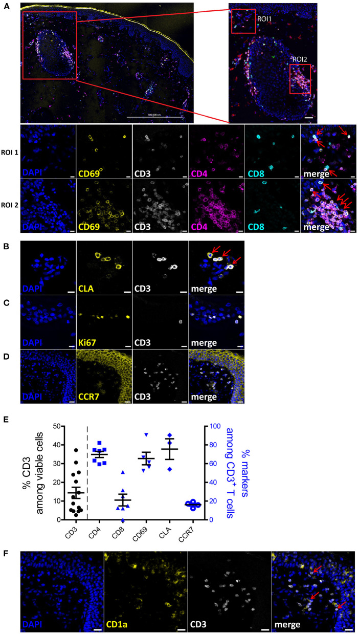
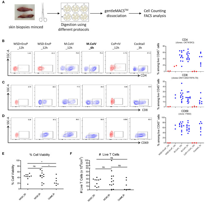
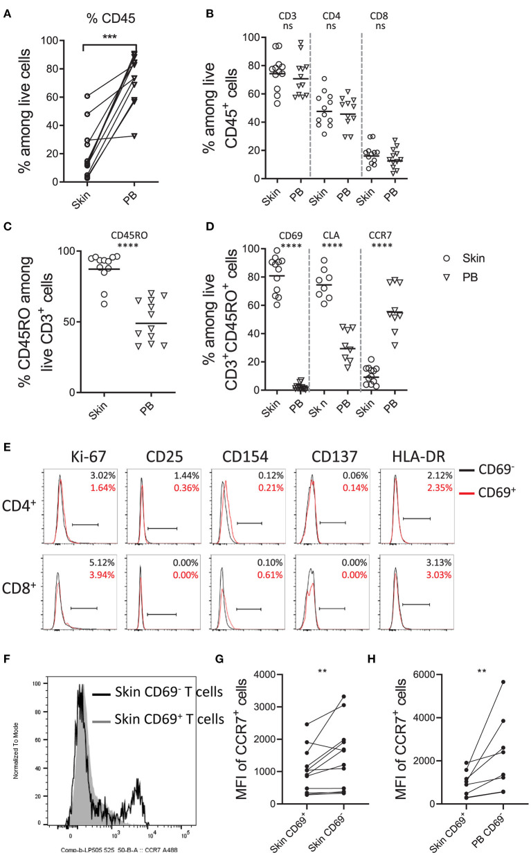
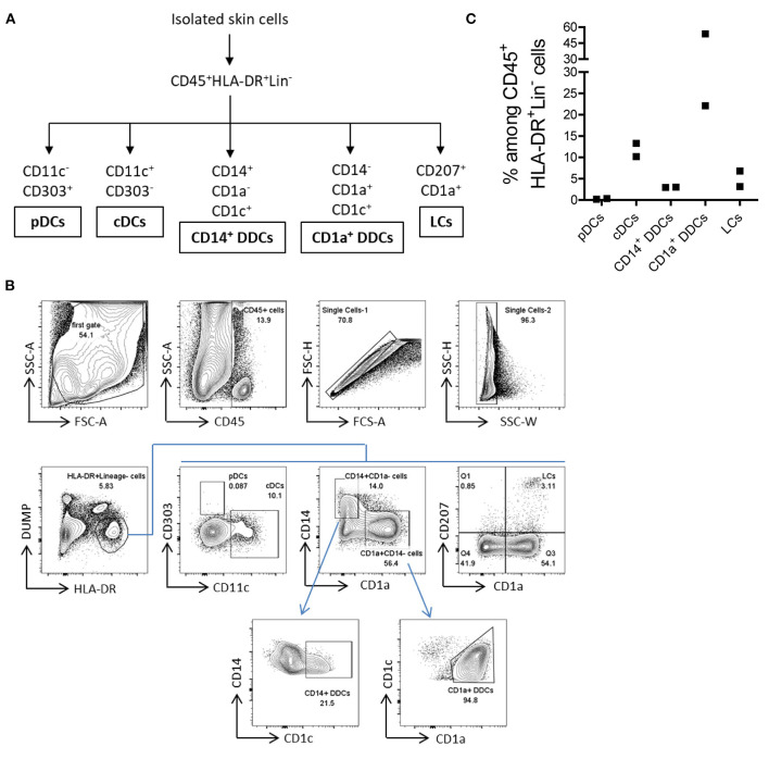
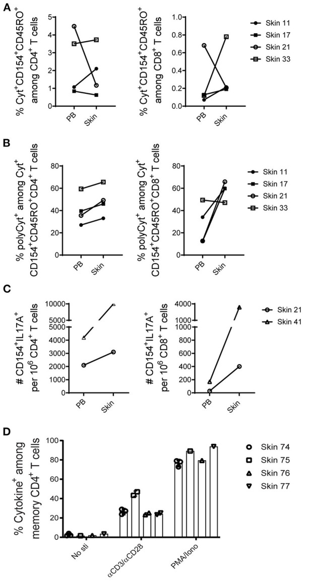
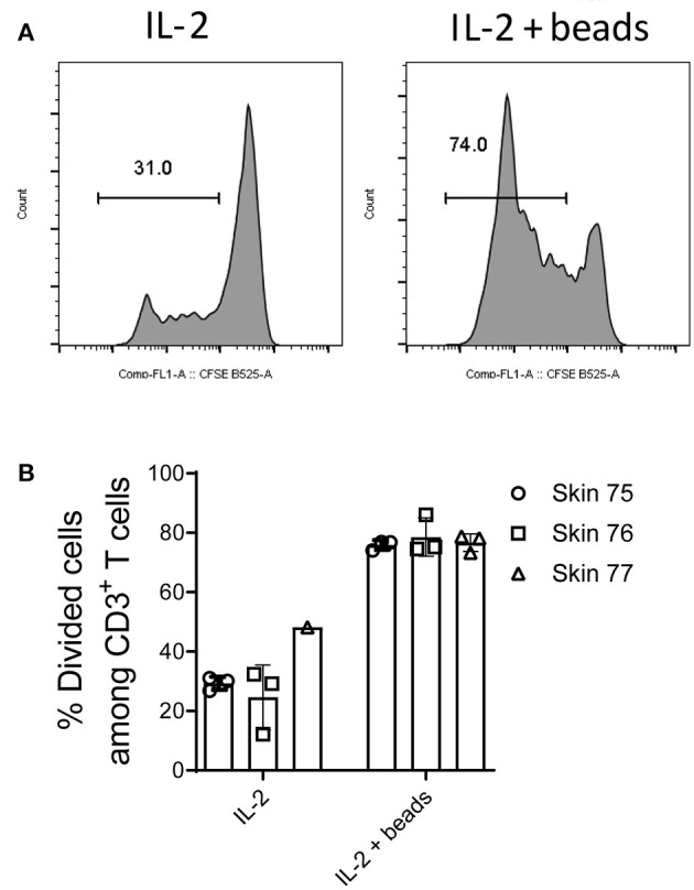
Studies in animal models have shown that skin tissue-resident memory T (TRM) cells provide enhanced and immediate effector function at the site of infection. However, analyses of skin TRM cells in humans have been hindered by the lack of an optimized isolation protocol. Here, we present a combinatorial strategy-the 6-h collagenase IV digestion and gentle tissue dissociation - for rapid and efficient isolation of skin TRM cells with skin tissue-specific immune features. In comparison with paired blood circulating memory T cells, these ex vivo isolated skin T cells express typical TRM cell markers and display higher polyfunctional properties. Moreover, these isolated cells can also be assessed for longer periods of time in ex vivo cultures. Thus, the optimized isolation protocol provides a valuable tool for further understanding of human skin TRM cells, especially for direct comparison with peripheral blood T cells at the same sample collection time.

Keywords: cell isolation; collagenase IV; epitope; gentle tissue dissociation; human skin; tissue-resident memory T cells; yield.


Conflict of interest statement

The authors declare that the research was conducted in the absence of any commercial or financial relationships that could be construed as a potential conflict of interest.

Figures

Figure 1
Figure 1
Phenotypic characterization of human skin cells in situ. (A) A 3 × 4 tile scan image of skin section stained with DAPI (blue), CD69 (yellow), CD3 (white), CD4 (violet) and CD8 (turquoise). Region of interest (ROI) 1 is a representative image of the cells located in the epidermis and ROI2 is a representative image of the cells around a hair follicle in the dermis. (B–D,F) Skin sections were stained with DAPI (blue) and CD3 (white) as well as one of the following: CLA (B), Ki-67 (C), CCR7 (D) or CD1a (F). Scale bar: 500 μm for (A) upper left, 20 μm for (A) upper right, (D,F); 10 μm for A-ROI1, A-ROI2, (B,C). Co-expression of CD3+CD4+/CD8+ and CD69+ cells, and CLA+ and CD3+ cells are indicated by red arrows. Representative image sets from three independent experiments are shown. Scale bar: 20 μm for (A,D,F); 10 μm for (A–C). (E) Frequencies of CD3+ T cells among total cells (left y-axis) and frequencies of indicated subpopulations of T cells among CD3+ T cells (right y-axis), according to image cell quantification (n = 3; 14 fields).
Figure 2
Figure 2
Modified Collagenase IV protocol best preserves the epitopes of surface antigens with high cell viability and yield. (A) Schematic workflow of isolating cells from human skin samples. (B–D) Frequencies of CD4+ (B), CD8+ (C) and CD69+ (D) T cells among live CD45+ lymphocytes isolated by six different isolation protocols: (1) WSD+EnzP_12 h (n = 2), (2) WSD-EnzP_12 h (n = 2), (3) M.CoIV_12 h (n = 12), (4) M.CoIV_6 h (n = 12), (5) CoP+/-CoIV_12 h (n = 3), and (6) Cocktail_3 h (n = 7). Each dot represents data obtained from one donor. Red dots showing cells isolated from skin samples using protocols that did not preserve the CD4 epitope. (E) Frequencies of viable cells and (F) the total number of viable T cells isolated by using the M.CoIV_12 h, M.CoIV_6 h and cocktail_3 h isolation protocols. Statistical significance was calculated by two-tailed, unpaired t-test with Welch's correction. p < 0.05 (*).
Figure 3
Figure 3
Phenotypic characterization of skin T cells by flow cytometry. Frequencies of CD45+ lymphocytes (A), CD3+, CD4+, and CD8+ T cells (B), CD45RO+ memory T cells (C), and CD69+, CLA+ and CCR7+ cells among memory CD3+ T lymphocytes (D) in paired (A) and unpaired human skin and peripheral blood samples (B–D). (E) Overlay of histograms showing the percentages of cells expressing proliferating and putative activation markers (Ki-67, CD25, CD154, CD137 and HLA-DR) on CD69 (black line) and CD69+ (red line) memory CD4+ and CD8+ T cells. Percentages are shown in the upper right of each plot. Representative data from more than 10 independent experiments are shown. (F) Overlay of histograms showing the expression of CCR7 on CD69+ (filled gray area) and CD69 (black line) skin T cells. Comparison of MFI (Mean Fluorescence Intensity) of CCR7+ cells between skin CD69+ and CD69 T cells (G) and between skin CD69+ and blood CD69 T cells. In (A,G,H), Wilcoxon matched-pairs signed rank test, two-tailed; in (B–D), unpaired T-test with Welch's correction, two-tailed. ****P < 0.0001, ***P < 0.001, **P < 0.005, ns.
Figure 4
Figure 4
Isolation of five major types of APCs from normal human skin by using the M. CoIV_6 h protocol. (A) Classification of five main types of APCs distinguished by their surface markers (22, 23). (B) Gating Strategy for analyzing APCs from ex vivo human skin cells. Lineage markers included CD3, CD20, CD34, and CD56. Dead cells were excluded by DAPI staining. pDCs and CDCs were distinguished by CD11c against CD303. CD14+ DDCs and CD1a+ DDCs were distinguished by CD1a against CD14 and further based on the CD1C expression. LCs cells were gated based on the expression of CD1a and CD207. (C) Frequencies of ex vivo pDCs, cDCs, CD14+ DDCs, CD1a+ DDCs and LCs among CD45+HLA-DR+Lin viable cells isolated from human eyelid skin samples. Representative data from two independent experiments are shown.
Figure 5
Figure 5
Functional capacities of T cells from skin and paired peripheral blood samples. (A–C), mononuclear cells isolated from five paired skin and PB samples were stimulated with the SEB, and the induced cytokine production (IFN-γ, IL-2, or TNF-α; alternatively IFN-γ, IL-2, TNF-α, or IL-17A) in memory CD4+ and CD8+ T cells was examined according to CD154 expression. For each subpopulation, the background (as detected in the anti-CD28 stimulated but otherwise equally treated control samples) was subtracted. (A) Antigen-specific CD154+cytokine+ (total cytokine-producing) memory CD4+ and CD8+ T cells are shown in frequencies among CD4+ and CD8+ T cells. (B) The proportions of polyCyt+-producing (more than one of the analyzed three or four cytokines TNF-α, IFN-γ, IL-2, or IL-17A) memory T cells among cytokine+CD154+CD45RO+ CD4+ and CD8+ T cells. (C) The absolute numbers of IL-17A+ cells per million CD4+ and CD8+ T cells isolated from two analyzed paired skin and blood samples upon SEB stimulation are shown. (D) Percentage of cytokine+ cells among skin memory CD4+ T cells in response to αCD3/αCD28 stimulation. No antigens and PMA/Ionomycin stimulation were included as controls. Bars with two or three data points are shown as the mean of replicates of cells analyzed from each sample.
Figure 6
Figure 6
Long-term cultures and expansion of ex vivo isolated skin T cells. Skin mononuclear cells were isolated using the optimized M.CoIV_6 h isolation protocol from three individual skin samples. (A) Representative histograms and (B) percentages of divided cells among skin CD3+ T cells cultured in medium supplemented with Proleukin (IL-2) (left) or in the presence of additional T cell expansion beads (right). In (B) bars with three data points represent the mean (± SEM) of three replicates of cells analyzed from each sample.

References

    1. Clark RA, Watanabe R, Teague JE, Schlapbach C, Tawa MC, Adams N, et al. . Skin effector memory T cells do not recirculate and provide immune protection in alemtuzumab-treated CTCL patients. Sci Transl Med. (2012) 4:117ra7. 10.1126/scitranslmed.3003008 - DOI - PMC - PubMed
    1. Gebhardt T, Wakim LM, Eidsmo L, Reading PC, Heath WR, Carbone FR. Memory T cells in nonlymphoid tissue that provide enhanced local immunity during infection with herpes simplex virus. Nat Immunol. (2009) 10:524–30. 10.1038/ni.1718 - DOI - PubMed
    1. Kumar BV, Ma W, Miron M, Granot T, Guyer RS, Carpenter DJ, et al. . Human tissue-resident memory T cells are defined by core transcriptional and functional signatures in lymphoid and mucosal sites. Cell Rep. (2017) 20:2921–34. 10.1016/j.celrep.2017.08.078 - DOI - PMC - PubMed
    1. Purwar R, Campbell J, Murphy G, Richards WG, Clark RA, Kupper TS. Resident memory T cells (T(RM)) are abundant in human lung: diversity, function, and antigen specificity. PLoS ONE. (2011) 6:e16245. 10.1371/journal.pone.0016245 - DOI - PMC - PubMed
    1. Stelma F, de Niet A, Sinnige MJ, van Dort KA, van Gisbergen K, Verheij J, et al. . Human intrahepatic CD69 + CD8+ T cells have a tissue resident memory T cell phenotype with reduced cytolytic capacity. Sci Rep. (2017) 7:6172. 10.1038/s41598-017-06352-3 - DOI - PMC - PubMed

Publication types

MeSH terms

LinkOut - more resources