. 2021 Feb;13(1):6900310.
doi: 10.1109/jphot.2020.3043685. Epub 2020 Dec 14.

Dual-Illumination Ultrasound/ Photoacoustic System for Cervical Cancer imaging

Affiliations

Dual-Illumination Ultrasound/ Photoacoustic System for Cervical Cancer imaging

Maryam Basij et al. IEEE Photonics J. 2021 Feb.

Abstract

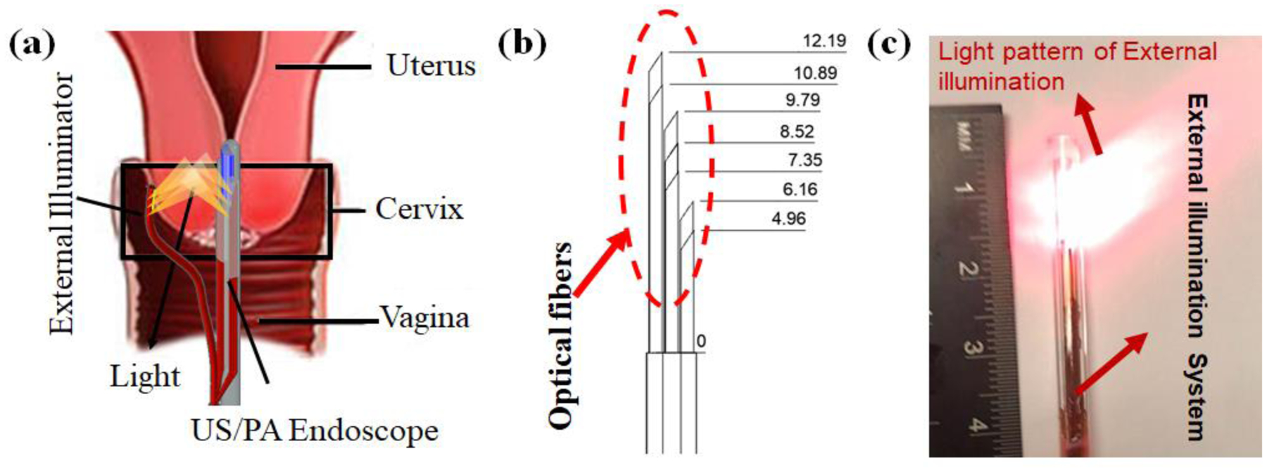
Early stage cancer detection technologies can provide functional information and potentially decrease the mortality rate caused by cervical cancer. In our previous work, a miniaturized ultrasound and photoacoustic endoscopic system has been developed to image the cervical tissue through the cervical canal to fulfills the need for a safe, low-cost, and high-resolution functional diagnostic system. However, the miniaturized size of endoscope and American National Standards Institute safety limits cause constraints of using high-intensity illumination during imaging. In addition, the strong light scattering of tissues limits the light penetration depth. Fortunately, the cervix anatomy allows for the delivery of additional light from the ectocervix by using an external illumination system. Here we propose a dual, co-planar illumination system, which can provide adequate illumination to the cervical tissue via combined internal and external light delivery strategies. Therefore, an increase in the area of light-tissue interaction allows us to raise the laser light energy while keeping fluence under safety limits. Thus, a reliable PA imaging can be obtained for the whole cervical tissue thickness. The system performance was tested using a Monte Carlo simulation, and laser-light fluence was calculated and compared at different depths within a simulated cervical-tissue model. The results indicated a higher and more uniform fluence in the Monte Carlo simulations. In addition, the photoacoustic imaging of the proposed system was evaluated by two cervical tissue-mimicking phantoms with human blood and graphite rods as inclusions inside it. In accordance with the simulations, the phantom study revealed a more reliable photoacoustic signal for the entire depth of the phantoms with an improved contrast to noise ratio and signal to noise ratio, and a higher coverage ratio of the imaging field of view. In summary, the dual-mode illumination system can provide more realistic information of inclusions within the tissue while considering safety limits, which can lead to more accuracy in biomarker detection for cervical cancer diagnostics.

Keywords: Dual illumination; Endoscope; Monte-Carlo; fiber optic; photoacoustic; side-firing; ultrasound.


Figures

Figure 1 :
Figure 1 :
A schematic of the dual-illumination US/PA endoscope-based imaging system for imaging the cervical tissue. When the integrated endoscopic imaging system passes through the cervical canal, the external light delivery system provides illumination through the ectocervix. (b) A schematic of our external illumination fiber equipped with seven optical fibers. The arrangement of the fibers is shown with their distance to the reference point on fiber bundle. (c) An image of the external light delivery system and its light pattern.
Figure 2:
Figure 2:
A schematic of the imaging system components for the dual-mode illumination US/PA endoscopic system. 680 nm pulse laser is used for generating PA images. A plano-convex lens is utilized for focusing the laser beam, and a beam splitter couples the pulsed laser light into the internal and external fiber bundles. The internal and external fibers are attached to each other before the distal end to ensure that the external light beam in positioned in front of the integrated US/PA endoscopic system.
Figure 3:
Figure 3:
A schematic of 3D design used for the Monte Carlo simulation of the dual-illumination imaging system. A cervical tissue with 30 mm thickness was irradiated by the external and internal illumination systems. A perfect absorber plane was placed at different depths to measure the light ray fluence.
Figure 4:
Figure 4:
A schematic of cervix mimicking phantoms made using porcine tissue embedded (a) with five US-transparent tubes filled with human blood with an interval distance of 5 mm (b) with twelve graphite rods with vertical and horizontal interval distance of 5 mm and 7.5 mm. The phantoms were imaged by applying three illumination techniques: (i) internal, (ii) external, and (iii) combined dual-mode illumination. The light pattern and US field of view are indicated by yellow and blue color, respectively.
Figure 5:
Figure 5:
Monte Carlo simulation of the light fluence for each illumination approach inside the cervical tissue with a thickness of 30 mm. (a-c) Simulated middle plane at 15 mm of each illumination strategy (d) Simulation of light pattern inside the tissue on fourteen sagittal planes with the increments of 0.2 mm along X-axis (e)The obtained light patten of internal and external illumination system on YZ plane by averaging the simulated light pattern over fourteen planes covering the elevational beam width of the US transducer (f) The mean and standard deviation of the incident light ray fluence of planes at depths of 7.5,15 and 22.5 mm from integrated US/PA endoscope. The results show how a higher and more consistent fluence can be obtained throughout the whole simulated cervical tissue thickness by using the dual-illumination system.
Figure 6:
Figure 6:
PA images of cervix mimicking tissue phantoms containing five blood filled tubes illuminated using three strategies: (a) internal, (b) external, and (c) dual-mode illumination. (d) PA amplitude, (e) the SNR and (f) the CNR of blood-filled tubes at different depths for each illumination method demonstrates the uniformity of the PA signal across the tissue with the combined dual-mode illumination system.
Figure 7:
Figure 7:
PA images of cervix mimicking tissue phantom containing twelve graphite rods illuminated using (a) internal, (b) external, and (c) combined dual-mode illumination systems. The results indicated that about 41%, 48%, and 91% of coverage percentage of tissue can be obtained from internal, external, and dual-illumination systems, respectively.

References

    1. Siegel RL, Miller KD, and Jemal A, “Cancer statistics, 2020,” Ca-a Cancer Journal for Clinicians, vol. 70, no. 1, pp. 7–30, January, 2020. - PubMed
    1. Cronin KA, Lake AJ, Scott S, Sherman RL, Noone AM, Howlader N, Henley SJ, Anderson RN, Firth AU, Ma J, Kohler BA, and Jemal A, “Annual Report to the Nation on the Status of Cancer, part I: National cancer statistics,” Cancer, vol. 124, no. 13, pp. 2785–2800, July 1, 2018. - PMC - PubMed
    1. Ward EM, Sherman RL, Henley SJ, Jemal A, Siegel DA, Feuer EJ, Firth AU, Kohler BA, Scott S, Ma J, Anderson RN, Benard V, and Cronin KA, “Annual Report to the Nation on the Status of Cancer, Featuring Cancer in Men and Women Age 20–49 Years,” J Natl Cancer Inst, vol. 111, no. 12, pp. 1279–1297, December 1, 2019. - PMC - PubMed
    1. Cohen PA, Jhingran A, Oaknin A, and Denny L, “Cervical cancer,” Lancet, vol. 393, no. 10167, pp. 169–182, January 12, 2019. - PubMed
    1. Saslow D, Runowicz CD, Solomon D, Moscicki AB, Smith RA, Eyre HJ, Cohen C, and American Cancer S., “American Cancer Society guideline for the early detection of cervical neoplasia and cancer,” CA Cancer J Clin, vol. 52, no. 6, pp. 342–62, Nov-Dec, 2002. - PubMed

LinkOut - more resources