. 2018 Nov 9;11(4):10.16910/jemr.11.4.6.
doi: 10.16910/jemr.11.4.6.

Comparison of the precision of smooth pursuit in humans and head unrestrained monkeys

Affiliations

Comparison of the precision of smooth pursuit in humans and head unrestrained monkeys

Jan Churan et al. J Eye Mov Res. .

Abstract

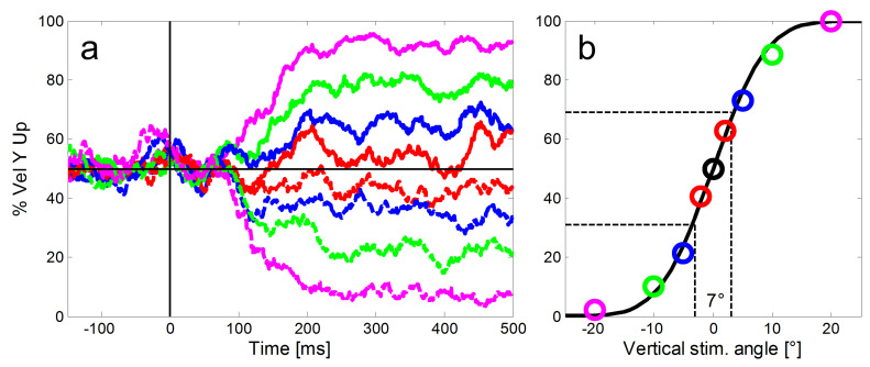
Direct comparison of results of humans and monkeys is often complicated by differences in experimental conditions. We replicated in head unrestrained macaques experiments of a recent study comparing human directional precision during smooth pursuit eye movements (SPEM) and saccades to moving targets (Braun & Gegenfurtner, 2016). Directional precision of human SPEM follows an exponential decay function reaching optimal values of 1.5°-3° within 300 ms after target motion onset, whereas precision of initial saccades to moving targets is slightly better. As in humans, we found general agreement in the devel-opment of directional precision of SPEM over time and in the differences between direc-tional precision of initial saccades and SPEM initiation. However, monkeys showed over-all lower precision in SPEM compared to humans. This was most likely due to differences in experimental conditions, such as in the stabilization of the head, which was by a chin and a head rest in human subjects and unrestrained in monkeys.

Keywords: Eye movement; eye tracking; head unrestrained; non-human primates; saccades; smooth pursuit.


Conflict of interest statement

The authors declare that the contents of the article are in agreement with the ethics described in http://biblio.unibe.ch/portale/elibrary/BOP/jemr/ethics.html and that there is no conflict of interest regarding the publication of this paper.

Figures

Figure 1:
Figure 1:
Experimental set-up. The mouthpiece (white) of the reward system and the photoelectric barrier (within the black block with the half-circular opening) to detect whether the monkey’s head was in a position suitable for the measurement of eye movements are in front, the monitor and the camera for eye movement measurements are in the back.
Figure 2:
Figure 2:
Diagram of the stimuli used to measure the directional precision of initial saccades and smooth pursuit (after Braun & Gegenfurtner, 2016). Left: In the Ramp paradigm, the eye movement target moves randomly after an initial fixation period left- or rightward at 10°/s. No or one out of eight different vertical components of +/- 20°, 10°, 5°, or 2° was added unpredictably to the horizontal ramp direction. Right: In the Step-Ramp paradigm after initial fixation the target first makes a step contraversive to the direction of the upcoming ramp motion in one of the indicated directions. Different colors represent different vertical components, solid lines represent upward- and dashed lines downward vertical components.
Figure 3:
Figure 3:
Illustration of our method to calculate the directional precision of SPEM in time bins of 1 ms. a: For the calculation of the directional precision we used all step-ramp trials collected from each monkey (here example data from monkey ME). For each ramp direction (shown here in different colors and line types as introduced in Figure 2), we determined the proportions of trials with an upward eye velocity (y-axis) relative to the horizontal baseline (black horizontal line). Shortly after 100 ms SPEM started to deviate according to the ramp direction of the target. b: In a second step a cumulative Gaussian was fitted to the proportions of upward eye movements to estimate the directional precision at each point in time (color codes of the single markers represent different vertical components of the stimulus as introduced in Figure 2). The slope of the function - quantified as the difference of vertical stimulus angles that was required to reach 31% and 69% upward responses (indicated by the dashed black lines) was used to measure the precision of the eye movements. Here the directional threshold was 7°.
Figure 4:
Figure 4:
Averaged eye velocities for the ramp (left column) and step-ramp (right column) paradigms for monkey MB. The different vertical stimulus components are coded by different line colors as shown in in Figure 2. Like in Figure 2, solid lines represent upward stimulus movements, while dashed lines represent downward stimulus movements the vertical components are 2° (red), 5° (blue), 10° (green) and 20° (magenta). a: Horizontal eye velocities, b: Vertical eye velocities, the vertical target velocities are indicated by thin horizontal lines in the respective color. c: Percentages of trials (y-axis) in which the vertical eye velocity was ‘upward’. All eye velocity traces are baseline corrected for the eye movement to pure horizontal target motion.
Figure 5:
Figure 5:
Averaged eye velocities for the ramp (left column) and step-ramp (right column) paradigms for monkey ME. All conventions like in Figure 4.
Figure 6:
Figure 6:
Time courses of direction thresholds of smooth pursuit and initial saccades for monkey MB (left) and ME (right) with respect to target motion onset. The direction thresholds for pure pursuit measured with the step-ramp paradigm are plotted in light blue and the fitted decay functions (Equation 1) in dark blue. Direction thresholds for initial saccades measured with the ramp paradigm are plotted in green for a 30 ms time window starting at saccade onset. This plot allows the comparison of the directional thresholds of saccades (green) and pursuit (STEP, orange) during the same time-window relative to the onset of target motion. For monkey ME, we marked in magenta the directional pursuit thresholds after additional 80 ms which corresponds to the saccadic reaction time of monkey MB. The average directional thresholds during steady-state SPEM (black lines) were calculated from 300 ms to 500 ms after the onset of stimulus motion.
Figure 7:
Figure 7:
a: Comparison of the normalized time courses of directional precision in the step-ramp paradigm for two monkeys (blue lines) and the average of four trained human subjects (black line, grey area shows std) from Braun & Gegenfurtner (30). The corresponding peri-saccadic precisions and saccadic latencies are shown as red lines for the NHPs (dashed: monkey ME; solid: monkey MB) and a green cross (showing the average latency of the saccades as well as the mean and std of peri-saccadic direction thresholds) for the human subjects. b: Direction thresholds during the initial saccade in the ramp-paradigm and the corresponding times during SPEM-onset in the step-ramp paradigm (STEP) for monkey MB and ME (red circles). Thresholds were normalized by the asymptotic pursuit threshold. For comparison, data from four trained human subjects (green squares) and six untrained human subjects (black crosses) from Braun & Gegenfurtner (30).

Similar articles

Cited by

References

    1. Amiez, C., & Petrides, M. (2009, October). Anatomical organization of the eye fields in the human and non-human primate frontal cortex. Progress in Neurobiology, 89(2), 220–230. 10.1016/j.pneurobio.2009.07.010 - DOI - PubMed
    1. Blanke, M., Harsch, L., Knöll, J., & Bremmer, F. (2010, December). Spatial perception during pursuit initiation. Vision Research, 50(24), 2714–2720. 10.1016/j.visres.2010.08.037 - DOI - PubMed
    1. Bourrelly, C., Quinet, J., Cavanagh, P., & Goffart, L. (2016, December 1). Learning the trajectory of a moving visual target and evolution of its tracking in the monkey. Journal of Neurophysiology, 116(6), 2739–2751. 10.1152/jn.00519.2016 - DOI - PMC - PubMed
    1. Brainard, D. H. (1997). The Psychophysics Toolbox. Spatial Vision, 10(4), 433–436. 10.1163/156856897X00357 - DOI - PubMed
    1. Braun, D. I., & Gegenfurtner, K. R. (2016, October 1). Dynamics of oculomotor direction discrimination. Journal of Vision (Charlottesville, Va.), 16(13), 4. 10.1167/16.13.4 - DOI - PubMed

LinkOut - more resources